|
|

Diabetic Patient with Macular Edema and Blood Pressure 200/95605 views49-year-old decreasing vision over the last year. OD is 20/80, OS 20/80. blood pressure which was 200/95.
|
|
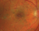
BDR and Diabetic Macular Edema with Fresh Laser Right Eye605 views60-year old man OD 20/60, OS 20/50 worsening vision both eyes
|
|

Angioid Streaks - New Extrafoveal Choroidal Neovascular Membrane Right Eye605 views61-year-old woman twelve days ago, saw a spot to the right of her central vision. OD is 20/25, OS is 20/25
Treated with one year of avastin with good result and no vision loss.
|
|

Central Retinal Artery Occlusion less than 24 hours - 69 year old man VA light perception605 views
|
|
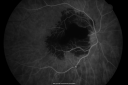
Ruptured Retinal Arterial Macroaneurysm - Submacular Hemorrhage - Vision 20/400 never recovered - Hypertension 205/94605 views
|
|

Ocular Ishemic Syndrome - Bilateral Carotid Occlusions - OD 20/60 OS 20/200 Enlarged Foveal Avascular Zone and Temporal Non-perfusion605 views75-year-old man had laryngeal cancer treated in 15 year ago with radiation. He recently was told that the carotid circulation is closed off on both sides. He does have a patent vertebral artery, which is supplying him pretty well and in addition to that his cardiac output is compromised by poor mitral valves. His consideration is to what can be done about that in the future.
VISUAL ACUITY: OD 20/60, OS 20/200
|
|

Basal Laminar Drusen - Cuticular Drusen - Equatorial Drusen - 70 Year Old Woman - OCT Map605 views70 year old woman has had macular drusen since her thirties and she said her sister has a similar problem. Recently the vision in the left eye has substantially declined. OD 20/16, OS 10/200.
|
|

Serous Retinal Detachment - Hypertensive Retinopathy - 165/120 - Indocyanine Green Angiogram605 views48-year-old woman noticed when she was blinking there was a white flash in the left eye starting about a month ago. She sees it temporally. OD 20/16, OS 20/20. There is a serous retinal detachment superotemporal to the optic nerve falling short of the fovea. Her blood pressure 165/120.
|
|

Punctate Inner Choroidopathy - Recurrent CNVM 4 years after initial treatment Left Eye - Indocyanine Green Angiogram605 viewsJune 7, 2012. This pleasant 35-year-old woman noticed decreasing vision in the left eye recently. She returns now noticing the vision in the left eye is declining. OD 20/50, OS 20/80. OS: Vertical C/D ratio is 0.3. There is a scar just inferonasal to the fovea, which has grown some towards the fovea with a little blood just under the fovea.
|
|

Haramotoma with ERM605 viewsHaramotoma with an ERM in the OS of a middle aged man
|
|

Non-specific (uncharaterized / unknown) Macular Dystrophy 605 views59-year-old man has macular dystrophy in both eyes. He had some vision changes in medical school in the 1980s and saw Dr. Gass for that. He had some pigment epithelial irregularities in both eyes. He had strabismus as a child and had muscle surgery. He is color blind, along with many people in his family, and as far as he knows, it is congenital.
20/25, 20/30
|
|

Primary Ocular Lymphoma - White Mass Left Eye - 112712 605 views
|
|

Reticular Macular Disease (Pseudo-drusen) Both Eyes - Occult CNVM and Macular Edema Right Eye 605 viewsSpectral Domain Optical Coherence Tomography
|
|
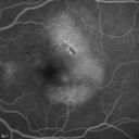
Polypoidal Choroidal Vasculopathy with Pulsating ICG polyp left eye605 views
|
|

Outer Retinal Holes - Probably Solar Retinopathy (history is negative)605 views
|
|

Choroideremia - Female Carrier - Patchy Atrophy of Choroid and RPE - Lyonization605 views
|
|

OCT shows lamellar macular hole left eye605 viewsWet AMD in right and better eye failed extend to 2 months but was able to stop therapy once he started on systemic Avastin for cancer. The last image shows the macula dry 9 months without local therapy. The left eye has a lamellar macular hole.
|
|

Central Areolar Choroidal Dystrophy605 viewsVision loss from early 60's. This 78 year old woman has choroidal atrophy centrally.
|
|

Syphilitic Chorioretinitis605 viewsSevere outer retinal atrophy and RPE non-geographic atrophy from syphilis. Vision improved from 20/63 to 20/32 with therapy. Images look a lot like DUSN. Patient had several surgeries (vitrectomy for floaters, vitrectomy for macular pucker, Avastin injections and macular laser for edema) prior by other retina specialists prior to being correctly diagnosed and treated.
|
|

CRVO with Paracentral Acute Middle Maculopath - PAMM605 views56 year old woman with sudden vision loss, hemorrhages in all four quadrants. FA shows good retinal circulation. The OCT shows PAMM lesions in the affected eye. Vision did improve on second visit from 20/160 to 20/60 in about a month
|
|

605 views
|
|

Subretinal Hemorrhage - Commotio Retinae - Macula - hit with baseball605 views15 year old with VA 20/60 3 days after being hit with baseball in the right eye
|
|

Multifocal Choroiditis - Linear Scars - Schlaegel lines605 views66 year old woman with macular pucker right eye. Images show typical linear peripheral scars for MFC or POHS - this is unilateral.
|
|

Fundus Albipunctatus605 views12 year old female with normal vision. She has 4 siblings all of whom have either white spots or spots on IR. Genetic testing by parents was deferred.
|
|

Choroidal Nevus with Drusen605 viewsLow risk choroidal nevus with many overlying drusen
|
|

Stargardt's Macular Dystrophy605 views62 year old man with good vision
|
|

Macular Pucker in eye following giant retinal tear RD repair post-op OCT604 viewsnow that the bubble is gone, but he is starting to notice distortion of central vision. He had a giant retinal tear retinal detachment repair emergently done on November 8, 2007, and subsequent laser on November 15th.
VISUAL ACUITY: Vision OD 20/400.
6 weeks post-op VA 20/40
|
|

Choroidal Melanoma Medium604 views33-year-old man with a history of decreased in the right eye. He had blurring in the eye about a year ago. He reports that it improved spontaneously until it was “nearly resolvedâ€. He said he had some persistent distortion, but that over the past couple of weeks he has noticed this dark spot recurring in the right eye, the left eye is unaffected. He does not have any flashes, floaters or change in his peripheral visual acuity. 20/80 OD, 20/16 OS. No improvement with pinhole. IOP: 12 OD, 11 OS.
POSTERIOR SEGMENT EXAMINATION: Shows a clear view with no PVD in either eye. C/D ratio is 0.2 OU. Large retinal vessels are unremarkable. The macula of the right eye has a brown choroidal lesion with pigment overlying the surface and orange spots along its lateral surface. The left macula is normal.
B-SCAN: B-scan ecography of the ultrasound of the right eye shows a mass about 5 mm high with a moderate to low internal reflectivity on A-scan echogram. There is serous fluid inferiorly on the echogram
|
|

Central Retinal Vein Occlusion and Cystoid Macular Edema - Acute - Vision 20/50 604 views68-year-old man six weeks of vision loss right eye from CRVO and CME, vision right eye 20/50
|
|

Hypertensive Retinopathy One Year later with good Vision IgA Nephropathy604 viewsOD 20/30, OS 20/25 One year following initial episode.
|
|

Best Disease for 30 Years - 20/50 Vision both eyes - 61 Year Old Woman604 views61-year-old woman was diagnosed with Best’s disease about 30 years ago. She and her many family members were tested, both at the VA Hospital and then at USF. OD 20/50, OS 20/50
|
|
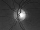
Optic Atrophy Left Eye and Benign Pituitary Tumor - VA 20/20 Right Eye 20/160 Left Eye604 views58-year-old man has had gradually declining vision in the left eye over about the last 18 months. Vision OD is 20/20, OS is 20/160
OCT SCAN: The OCT scan of the nerve fiber layer in the right eye shows a thickness of 88 microns, the left eye 59 microns.
MRI did show a suprasellar mass which is affecting the optic chiasm and is responsible for the vision loss. The tumor was resected and was a benign pituitary tumor.
|
|

Basal Laminar (cuticular) Drusen both eyes 60 Year Old Woman604 views60-year-old woman with minimal vision loss. Vision OD is 20/30 OS 20/40
|
|

Hyperpigmentation of the Retinal Pigment Epithelium in Dry AMD604 views87-year-old woman has wet age-related macular degeneration in the left eye. Vision OD is 20/25, OS is 20/400
|
|

Non-Ischemic Central Retinal Vein Occlusion in Young Man (30 years) - Many Cotton Wool Spots604 views30-year-old man with a history of a central retinal vein occlusion for which he was first examined on 2 months ago and his vision then was 4/200. He responded nicely to two Avastin treatments. OD is 20/20, OS is 20/60
|
|
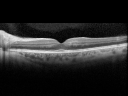
Juxtafoveal Retinal Telangiectasia (Telangiectasis) - MacTel - Striking Gray Ring in Macula - SDOCT604 views59-year-old woman decreasing vision in the right eye for a few years. She has had diabetes for five years. OD 20/100, Pinhole 20/40. OS 20/20
|
|

Retinal Detachment604 views
|
|
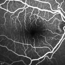
Polypoidal Choroidal Vasculopathy with Pulsating ICG polyp left eye604 views
|
|

Presumed Ocular Histoplasmosis with new CNVM in right and better eye - VA 20/80 OD; 20/800 OS604 viewsPeripheral punched out small scars typical for OHS
|
|

Presumed Ocular Histoplasmosis with new CNVM in right and better eye - VA 20/80 OD; 20/800 OS604 viewsFundus Autofluorescence shows scar as dark
|
|

Trousseau Sign - CRVO and Pancreatic Cancer604 viewsLeft eye shows CRVO with severe edema. The right eye has multiple small hemorrhages. Some of this is probably anemic retinopathy and some from hypercoaguability of cancer. The patient has pancreatic cancer.
|
|

Syphilitic Chorioretinitis604 viewsSevere outer retinal atrophy and RPE non-geographic atrophy from syphilis. Vision improved from 20/63 to 20/32 with therapy. Images look a lot like DUSN. Patient had several surgeries (vitrectomy for floaters, vitrectomy for macular pucker, Avastin injections and macular laser for edema) prior by other retina specialists prior to being correctly diagnosed and treated.
|
|
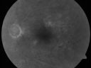
Clinically Significant Diabetic Macular Edema both Eyes with Exudates603 views56-year-old woman with diabetes for twelve years. She has noticed blurring of the vision in the right eye for about six months. OD is 20/70, OS is 20/25.
|
|

Acute Central Serous Chorioretinopathy with Symptoms in Hazy Vision Both Eyes603 views48-year-old man with hazy vision in the right eye for about three days. He has a little more of a headache than usual. He is under a fair amount of stress. He does not take any steroids that he knows of. He does use a nasal spray, which he is not sure if that is a steroid spray or not. He is going to call us with that medicine. I checked his blood pressure today is was 115/95.
VISUAL ACUITY: OD 20/15, OS 20/25
|
|
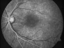
Juxtafoveal Telangiectasis - After Hemorrhage Absorbed Right Eye with Avastin Injections603 views73-year-old woman has juxtafoveal retinal telangiectasis in both eyes. The right eye has stage 5 disease. Her last Avastin treatment in the right eye was in 6 months ago. The right eye has been the worst of her two eyes for years.
VISUAL ACUITY: Vision OD is 20/200, OS is 20/50.
|
|
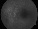
Non-Arteritic Anterior Ischemic Optic Neuropathy - Peripapillary Choroidal Ischemia603 views68-year-old woman noticed central vision loss in the right eye about a week ago. It is getting a little bit better. OD 20/160, OS 20/25.
|
|
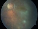
Chronic Macula-off Tractional Retinal Detachment Left Eye Proliferative Diabetic Retinopathy603 views81-year-old man with diabetes for 24 years has proliferative diabetic retinopathy in both eyes with a chronic tractional retinal detachment in the left eye 4 years old. OD is 20/40, OS is 2/200
|
|

Wet Age-related Macular Degeneration (AMD) Radiating Chorioretinal Folds from Contracting Macular Scar603 views93-year-old man has wet age-related macular degeneration in his left eye with a subfoveal pigment epithelial detachment.
Vision OS is 20/40. There are striking radiating chorioretinal folds emanating off of the scar, which suggests it is contracting.
|
|
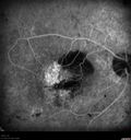
Post-Retinal Detachment Repair Occult Subfoveal CNVM603 viewsThis pleasant 56-year-old man has had a complex retinal detachment repair in the right eye. I most recently removed his silicone oil 3 months ago. His vision now is the best it has ever been.
VISUAL ACUITY: OD 4/200
|
|

Choroidal Melanoma, Post op Radioactive Implant603 views50-year old male who had a Radioactive Implant for Choroidal Melanoma in the left eye. VA is 20/150. VA has remained the same for the last 3-months.
|
|
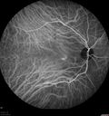
Serous Retinal Detachment - Hypertensive Retinopathy - 165/120 - Indocyanine Green Angiogram603 views48-year-old woman noticed when she was blinking there was a white flash in the left eye starting about a month ago. She sees it temporally. OD 20/16, OS 20/20. There is a serous retinal detachment superotemporal to the optic nerve falling short of the fovea. Her blood pressure 165/120.
|
|

Corneal Abrasion with Foreign Body Present 603 viewsER patient comes in with corneal abrasion in the left eye which was getting worst. VA 20/25. Slit lamp exam showed corneal abrasion superiorly at 2-o'clock. Flipped eyelid and foreign body appeared on the upper tarsal plate. Foreign body was removed.
|
|

Peripheral Retinal Degeneration603 viewsPatient comes in with double vision. VA was 20/20 in both eyes. Fundus exam shows Retinal Degenerative changes in both eyes. Offer to correct double vision with temporary Fresnel prism
|
|

Hypertensive Retinopathy603 viewsPatient comes in complaining of spots in vision in both eyes. VA was 20/25 - right eye and 20/20- left eye. Fundus exam reveals little hemorrhages with cotton wool spots due to hypertension and anemia.
|
|

Cytomegalovirus Retinitis603 viewsPatient who is HIV Positive with Cytomegalovirus Retinitis. Patient was treated with Gangcyclovir Intra-Vitreal injection.
|
|

Papilledema - Sagittal Cerebral Vein Thrombosis - Lung Transplant Cystic Fibrosis 36 Year Old Man603 viewsColor Photo
|
|
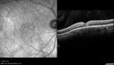
Macular Telangiectasia - Stage 3 right eye 20/40 vision603 viewsSpectral Domain OCT Line Scan
|
|

Choroideremia - Female Carrier - Patchy Atrophy of Choroid and RPE - Lyonization603 views
|
|

Hairy Cell Leukemia - Retinal Hemorrhage and twig Branch Vein Occlusion603 views79 year old man He has had hairy cell leukemia since 2002. He is in remission. His last blood tests were 9/2018. He just moved down here and needs a new leukemia doctor. His vision is fine.
VA OD: Dcc20/25
VA OS: Dcc20/25
IOP: TP: OD:12 OS:12
|
|

Syphilitic Chorioretinitis603 viewsSevere outer retinal atrophy and RPE non-geographic atrophy from syphilis. Vision improved from 20/63 to 20/32 with therapy. Images look a lot like DUSN. Patient had several surgeries (vitrectomy for floaters, vitrectomy for macular pucker, Avastin injections and macular laser for edema) prior by other retina specialists prior to being correctly diagnosed and treated.
|
|

Cystoid Macular Edema - Xalatan - ECCE was done 20 years previously602 views71 Year old woman who noticed about a month ago the central vision in the right eye fogged up. OD 20/200 POSSIBLE XALATAN INDUCED CYSTOID MACULAR EDEMA
|
|

Juxtafoveal Telangiectasis and Newly Decreasing Vision602 views70-year-old woman followed for 10 years with IJFT with newly decreasing vision
|
|

Chronic Pseudophakic Cystoid Macular Edema in a Patient with Diabetes602 views74-year-old woman has chronic cystoid macular edema in the left eye. She has had vision loss intermittently in that eye for 2 years, the right eye, unfortunately, lost vision from a central retinal vein occlusion in 6 years ago.
|
|
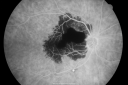
Ruptured Retinal Arterial Macroaneurysm - Submacular Hemorrhage - Vision 20/400 never recovered - Hypertension 205/94602 views
|
|

Retina Attached Under Oil after oil exchange, perfluoron, laser602 views
|
|

Choriodal Neovascular Membrane - Subfoveal - Pseudo-Retinitis Pigmentosa - 50 years post Retinal Detachment Repair602 views86-year-old with a history of decreased vision in the right eye. The patient reports she has lost vision in the right eye over the past two months or so. She reports the vision was always compromised in that eye since retinal detachment surgery 50 years ago with a scleral buckler. Vision dropped from 20/50 to 20/200 right eye in 2 months.
|
|

Plaquenil (Hydroxycholorquine) Toxicity - Bull's Eye Maculopathy - 73 year old womand 25 Years therapy - 64 inches tall - 110 lbs602 views73-year-old woman has been on Plaquenil for twenty five years. She noticed loss of paracentral vision a few months ago. VISUAL ACUITY: OD 20/70, OS 20/30. Her color vision was 0 out 10 correct in both eyes and she also missed the controls.
|
|

Juxtafoveal Telangiectasis - After Hemorrhage Absorbed Right Eye with Avastin Injections602 views73-year-old woman has juxtafoveal retinal telangiectasis in both eyes. The right eye has stage 5 disease. Her last Avastin treatment in the right eye was in 6 months ago. The right eye has been the worst of her two eyes for years.
VISUAL ACUITY: Vision OD is 20/200, OS is 20/50.
|
|

Nanopthalmous (Microphthalmous) Chorioretinal Folds and Choroidal Thickening Both Eyes602 views67-year-old woman with microphthalmos/nanophthalmos Her vision has gradually been getting a little bit worse, especially in the right eye. Also the anterior chamber is getting crowded and there is concern about the need for cataract surgery. She has one sister and one cousin who also have nanophthalmos.
VISUAL ACUITY: OD 20/50, OS 20/80.
|
|

Pattern Dystrophy - Adult Vitelliform Macular Degeneration602 views72-year-old woman 2 months post ECCE with poor vision since surgery especially left eye.
VISUAL ACUITY: OD 20/50, OS 20/200
|
|

Retinitis Pigmentosa Newly Diagnosed 55 Year Old602 views55-year-old noticed his vision declining recently, over the last few weeks, particularly in the right eye. He said previous to that the vision was not normal but was pretty good. OD is 20/100, OS is 20/50
OCT SCAN: There is macular thickening in both eyes with intraretinal cysts.
|
|

Retained Lens Fragments and Dislocated Intraocular Lens one week following complicated cataract surgery602 views78 year old female vision is 20/200. She developed a tear and RD during surgery for the lens fragments and has since developed PVR and had 5 surgeries. Her vision is now hand motion.
|
|

Vitreomacular traction - recent vision loss Right Eye - initially worse after Jetrea602 views
|
|

Acute AION OS - Mild disc pallor OD602 viewsSudden vision loss OS x 1 week with optic nerve swelling. Evidence of optic atrophy in right eye.
|
|

Diabetic Retimopathy with Peripherial Ischemia602 views30 year old woman with decreased vision right eye
|
|

Hairy Cell Leukemia - Retinal Hemorrhage and twig Branch Vein Occlusion602 views79 year old man He has had hairy cell leukemia since 2002. He is in remission. His last blood tests were 9/2018. He just moved down here and needs a new leukemia doctor. His vision is fine.
VA OD: Dcc20/25
VA OS: Dcc20/25
IOP: TP: OD:12 OS:12
|
|

Macular Telangiectasia and dry AMD - Simulating wet AMD602 viewsThis patient was told different things by different retina specialists. Her case is complex because she has OCT findings of Macular Telangiectasis and dry AMD. This makes it look like she has wet AMD. Her vision is stable.
|
|

Pattern Dystrophy - Probably Early Butterfly602 views33 year old with normal vision and no visual complaints - normal color vision
|
|

Choroidal Nevus - Macula - Flat602 views37 year old female with 5 year stable nevus Left eye - OCT shows flat nevus (EDI scan)
|
|

Myopic Degeneration - Inferior staphyloma with severe atrophy602 views76 year old with gradual vision loss. 20/32 OD; 20/40 OS
|
|

Diabetic Patient with Macular Edema and Blood Pressure 200/95601 views49-year-old decreasing vision over the last year. OD is 20/80, OS 20/80. blood pressure which was 200/95.
|
|

Diabetic Patient with Macular Edema and Blood Pressure 200/95601 views49-year-old decreasing vision over the last year. OD is 20/80, OS 20/80. blood pressure which was 200/95.
|
|

Toxoplasmosis - Inner Retinal - Recurrent - Treated with Bactrim DS 601 views43-year-old man has macular toxoplasmosis. He has responded nicely to Bactrim OS: 20/60
|
|

New BRVO Left Eye Old BRVO Right Eye - Left Eye Shows Twig Superior Vein Occlusion with Edema Arrow Shows Site of Occlusion601 views90-year-old man decreased vision maybe one year since cataract surgery he is no longer able to read. OD 20/200, OS 20/200
|
|

Juxtafoveal Telangiectasis - (MacTel) - Stage 4 both Eyes601 views57-year-old woman with a history of decreased vision. The patient reports that her vision has been gradually decreasing in the left more so than the right eye. 20/50 OD and 20/60
|
|

Acute Central Serous Chorioretinopathy with Symptoms in Hazy Vision Both Eyes601 views
|
|

Proliferative Diabetic Retinopathy in patient with Moyamoya Disease601 views28-year-old womandiabetic since age nine, and she also has had multiple strokes. OD is 20/30, OS is 20/30.
|
|

Indocyanine Green Angiogram - Enhanced S Cone Syndrome - Goldmann Favre - 601 views55-year-old woman while in college her vision was poor even with glasses and she sought evaluation for that. She was told after she had an electroretinogram at USF 15 years ago, that she had something with her blue cones. She does have poor night vision, but her reading vision is pretty good.
VISUAL ACUITY: OD 20/40, OS 20/40
|
|

Post Macula Retinal Detachment Repair On Pneumatic Retinopexy and subsequent Scleral Buckle Vitreous Opacity (No Vitrectomy)601 views55-year-old man had a scleral buckle for a macula-threatening retinal detachment in the left eye one month ago and a previous pneumatic retinopexy. Patient sees a big floater. His vision is improving.
VISUAL ACUITY: Vision OS is 20/50
|
|
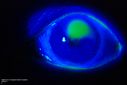
Cornea Ulcer/Abrasion601 views
|
|

Retinal Pigment Epithelial Detachment with no Sub-Retinal Fluid...601 viewsA 38-year old male who comes in with blurred vision in the left eye. VA is 20/30. Notices a defect inferior of his central vision. Did an Fluorescence Angiogram to determine an RPE with no sub retinal fluid. Also OCT confirms. Patient was injected with Avastin.
|
|

Lumigan (Bimatoprost) Cystoid Macular Edema - Eye Better 1 month after Stopping Lumigan601 views 82-year-old man had cataract surgery in 1993. He has had glaucoma for seven or eight years for a long time. He is taking Lumigan only in the left eye. About a year ago he started in the right eye and then starting just about a few months ago he has noticed intermittent blurring in the vision in the right eye. Vision 20/25 OD, 20/20 OS
|
|

Asymptomatic Juxtapapillary Choroidal Neovascular Membrane - Reticular Macular Disease (Wet AMD) 601 views
|
|

601 views
|
|
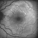
Polypoidal Choroidal Vasculopathy with Pulsating ICG polyp left eye601 views
|
|

Unilateral Acute Idiopathic Maculopathy (UAIM)601 views41 year old with serous retinal detachment OS and sudden vision loss. He had a viral prodrome. Coxscackie IgG was high but no acute infection was documented. Vision improved in about 2 months without therapy. - Right eye has spot nasal to the optic nerve
|
|

Syphilitic Chorioretinitis601 viewsSevere outer retinal atrophy and RPE non-geographic atrophy from syphilis. Vision improved from 20/63 to 20/32 with therapy. Images look a lot like DUSN. Patient had several surgeries (vitrectomy for floaters, vitrectomy for macular pucker, Avastin injections and macular laser for edema) prior by other retina specialists prior to being correctly diagnosed and treated.
|
|

Late Onset Retinal Degeneration (L-ORD)601 views55 year old with acute vision loss from a CNVM in the right eye. He responded to Lucentis therapy. His mother and her family has been confirmed genetically to have L-ORD and were part of the early reports.
|
|

Scleromalacia and Uveitis601 viewsPoor vision from uveitis and cataract. She was treated with Remicade and the uveitis abated and she had the right eye cataract done. The initial vision was 20/400 OD, 20/200 OS Final vision 20/60 OD, 20/32 OS
|
|

Branch Retinal Arterial Occlusion Right Eye - Atheromata - Both Eyes601 views83 year old woman with no symptoms - VA 20/40 in both eyes. Right eye has ST BRAO with silver wiring. Left eye has superotemporal atheromata
|
|
| 18050 files on 181 page(s) |
 |
|
|
|
|
48 | |
|
|
|
|


