|
|

Serous Retinal Detachment - Hypertensive Retinopathy - 165/120 600 views48-year-old woman noticed when she was blinking there was a white flash in the left eye starting about a month ago. She sees it temporally. OD 20/16, OS 20/20. There is a serous retinal detachment superotemporal to the optic nerve falling short of the fovea. Her blood pressure 165/120.
|
|

Punctate Inner Choroidopathy - Recurrent CNVM 4 years after initial treatment Left Eye - Fluorescein Angiogram600 viewsJune 7, 2012. This pleasant 35-year-old woman noticed decreasing vision in the left eye recently. She returns now noticing the vision in the left eye is declining. OD 20/50, OS 20/80. OS: Vertical C/D ratio is 0.3. There is a scar just inferonasal to the fovea, which has grown some towards the fovea with a little blood just under the fovea.
|
|

55 degree fa mid. od600 viewssimilar photo to autofluorescence
|
|
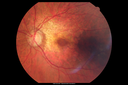
Posterior Polar Annular Choroidal Dystrophy600 views68 year old woman with know macular irregularities for over 20 years. She has since been followed another 6 years and her vision remains 20/25.
|
|

Rubeosis after Silicone Oil Removal Complex RD Repair with Peripheral 180 Degree Retinotomy (Inferiorly)600 views
|
|

Heidelberg False Color600 views
|
|

Optic Neuritis/Neuroretinitis600 viewsPt. states lost some vision in his left eye last week, was seen by eye MD and referred for eval. Also complains of pain in left eye with eye movement. VA was 20/400 in the left eye. Fundus photo and HD-OCT imaging show optic nerve swelling and fluid underneath the retina. Will consult with Neuro-Ophthalmologist for further evaluation.
|
|

Toxoplasmosis Retinitis - Vitritis and Anerior Uveitis600 views
|
|

Optic Disk Drusen - Pucker - Pattern Dystrophy600 views
|
|

Cystoid macular edema 4 months following cataract surgery with serous retinal detachment600 viewsCME which is chronic will often cause a serous reitnal detachment because of loss of the integrity of the external limiting membrane
|
|

Syphillitic Optic Neuropathy and Maculopathy600 views46 year old woman with 2 weeks of vision loss OD. VA 20/200 OD; 20/20 OS. there are posterior vitreous cells on OCT in both eyes. ICG, FAF, and FA show an abnormal macula OD. VA improved to 20/30 within a month with treatment with PCN.
|
|

Pattern Dystrophy - Probably Early Butterfly600 views33 year old with normal vision and no visual complaints - normal color vision
|
|

CRVO with Paracentral Acute Middle Maculopath - PAMM600 views56 year old woman with sudden vision loss, hemorrhages in all four quadrants. FA shows good retinal circulation. The OCT shows PAMM lesions in the affected eye. Vision did improve on second visit from 20/160 to 20/60 in about a month
|
|

Optic Nerve Pit Maculopathy600 views10 year old child who had inner retinal fenestration (see youtube video on scohen125 channel) and did very well.
|
|
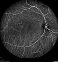
Central Serous Chorioretinopathy - Moderate600 views49 year old man with episodes in 2011, 2017, and 2018 - now with some paracentral vision loss in the right eye and subretinal fluid.
|
|

Torpedo Maculopathy - Left Eye600 views25 year old man with normal vision. Left macula has torpedo lesion
|
|

Diabetic Patient with Macular Edema and Blood Pressure 200/95599 views49-year-old decreasing vision over the last year. OD is 20/80, OS 20/80. blood pressure which was 200/95.
|
|
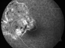
Serpiginous choroiditis599 viewsInactive scars in both eyes
|
|

Retinal Arterial Atheromata599 views68-year-old woman with records going back 7 years, which clearly document retinal arterial plaques in both eyes.
|
|

Birdshot Chorioretinitis - Retinal Vasculitis - HLA A29 Positive 599 views64-year-old woman three or four year’s fluctuations in her vision. She mostly notices them in bright light her vision glares out and she has difficulty seeing. PMHx: malaria in 1948 that was treated. She was told at one point she could possibly have had Familial Mediterranean fever.
She does intermittently get rashes around her arms. She has arthritis in her knees and ankles.
VISUAL ACUITY: OD 20/40, OS 20/50 - HLA - A29 Positive
|
|

599 views
|
|

Nanopthalmous (Microphthalmous) Chorioretinal Folds and Choroidal Thickening Both Eyes599 views67-year-old woman with microphthalmos/nanophthalmos Her vision has gradually been getting a little bit worse, especially in the right eye. Also the anterior chamber is getting crowded and there is concern about the need for cataract surgery. She has one sister and one cousin who also have nanophthalmos.
VISUAL ACUITY: OD 20/50, OS 20/80.
|
|

Concentric Annular Macular Dystrophy 4/200 2/200 vision 61 Year Old Macular Crystals599 views61-year-old woman has concentric annular macular dystrophy. She has had vision loss for 10 years who is no longer able to function even with the magnifiers and vision aids that she has. Her brother who is ten years younger than her, is recently diagnosed with the same macular dystrophy and her father and grandfather may have had similar problems in the past.
VISUAL ACUITY: OD 4/200, OS 2/200.
|
|

Pattern Dystrophy - Adult Vitelliform Dystrophy - Macular Degeneration599 views 78 Year Old Woman with vision has diminished in the right eye over the past couple of years, but the diminishment has been slow.
20/60+1 OD, 20/25 OS
|
|

Indocyanine Green Angiogram - Enhanced S Cone Syndrome - Goldmann Favre - 599 views55-year-old woman while in college her vision was poor even with glasses and she sought evaluation for that. She was told after she had an electroretinogram at USF 15 years ago, that she had something with her blue cones. She does have poor night vision, but her reading vision is pretty good.
VISUAL ACUITY: OD 20/40, OS 20/40
|
|
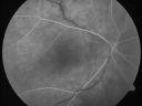
ECTOPIC DISCIFORM SCAR/PERIPHERAL CHOROIDAL NEOVASCULAR MEMBRANE – EXUDATE –INFLAMMATION IN REACTION TO THE PERIPHERAL VASCULAR LESION 599 views53-year-old woman has noticed some blurred vision in the right eye for a few years. In retrospect, she thinks it is possible since she was a teenager the right eye was always the weaker eye and that the left eye has always been the better eye. VISUAL ACUITY: Vision OD is 20/400
|
|

Pigmented Choroidal Mass Right Eye - Melanoma vs. Nevus vs. Unilateral Melanocytic Proliferation - Initial Visit599 views71-year-old man is vision has been bothering him for about a month. He does not notice one eye particularly worse than the other. OD is 20/40, OS is 20/25 - Initial Visit
|
|

Pigmented Choroidal Mass Right Eye - Melanoma vs. Nevus vs. Unilateral Melanocytic Proliferation - Initial Visit599 views71-year-old man is vision has been bothering him for about a month. He does not notice one eye particularly worse than the other. OD is 20/40, OS is 20/25 - Initial Visit
|
|

BESTS Disease599 views
|
|

Punctate Inner Choroidopathy - Recurrent CNVM 4 years after initial treatment Left Eye - Indocyanine Green Angiogram599 viewsJune 7, 2012. This pleasant 35-year-old woman noticed decreasing vision in the left eye recently. She returns now noticing the vision in the left eye is declining. OD 20/50, OS 20/80. OS: Vertical C/D ratio is 0.3. There is a scar just inferonasal to the fovea, which has grown some towards the fovea with a little blood just under the fovea.
|
|

Punctate Inner Choroidopathy - Recurrent CNVM 4 years after initial treatment Left Eye - Indocyanine Green Angiogram599 viewsJune 7, 2012. This pleasant 35-year-old woman noticed decreasing vision in the left eye recently. She returns now noticing the vision in the left eye is declining. OD 20/50, OS 20/80. OS: Vertical C/D ratio is 0.3. There is a scar just inferonasal to the fovea, which has grown some towards the fovea with a little blood just under the fovea.
|
|

Wet AMD - New Small Hemorrhage Both Eyes - Mature CNVM visible on Early Indocyanine Green Angiogram599 views
|
|

Reticular Macular Disease (Pseudo-drusen) Both Eyes - Occult CNVM and Macular Edema Right Eye 599 viewsIndocyanine Green Angiogram
|
|
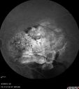
Idiopathic Polypoidal Choroidal Vasculopathy - Fluorescein Angiogram599 views
|
|

Choroideremia - Female Carrier - Patchy Atrophy of Choroid and RPE - Lyonization599 views
|
|
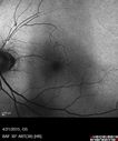
FAF shows increased FAF superiorly where the retina has been damaged because of loss of chromophores599 views
|
|

Choroidal Folds Hyperope (+5.00)599 views
|
|

Stargardt's Macular Dystrophy ABCA4 positive599 viewsImages show some progression over 3 years of macular dystrophy.
|
|

Syphilitic Chorioretinitis599 viewsSevere outer retinal atrophy and RPE non-geographic atrophy from syphilis. Vision improved from 20/63 to 20/32 with therapy. Images look a lot like DUSN. Patient had several surgeries (vitrectomy for floaters, vitrectomy for macular pucker, Avastin injections and macular laser for edema) prior by other retina specialists prior to being correctly diagnosed and treated.
|
|

Confluent Soft Drusen with Subetinal Fluid - Right Eye599 viewsAsymptomatic 85 year old man. OCT shows pocket of subfoveal fluid in the center of the macula. This could be mechanical from the drusen preventing the retina from settling down. Testing did not reveal a CNVM and subsequent OCT 6 weeks later showed no change in structure.
|
|

Recurrent MEWDS Left Eye - Teenage Girl599 viewsPatient presented in 2014 (first images) 2 months following vision loss left eye. Vision went from 20/60 - 20/20. OCT showed outer retinal irregularities which normalized. Then 3 years later in 2017 she had full blown MEWDS. She presented acutely and all the images are typical. Both episodes where in the same time of year, about 3 months after her flu shot for sports.
|
|

Choroidal Osteoma 13 Year Old599 views13 year old with 6 months of vision loss right eye and vision of 20/40
|
|

Retinal Capillary Hemangioma - Endophytic and Exophytic599 views46 year old female with normal vision - her father died of pancreatic cancer
|
|
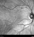
Occult Macular Dystrophy - 9 Years later 599 views53 year old man. Vision is better at 20/50 OU
|
|
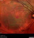
Lung Cancer Metastasis to Right Eye599 views63 year old female with vision loss for 6 weeks. She had been scheduled for cataract surgery and then a tumor was found. She was ultimately diagnosed with lung cancer. Vision is 20/100
|
|

Adult Pseudovitelliform Macular Dystrophy599 views89 year old female states dark smudge on ceiling during night time came and went. Decreased vision in the left eye
VA OD: Dcc20/32
VA OS: Dcc20/63
|
|

Wet AMD 62 year old male smoker 598 views62-year-old man has wet age-related macular degeneration in the right eye. He has been undergoing treatment with Lucentis for that. Then his left eye developed hazy vision a few weeks ago while he was overseas and now his vision is hazy in both eyes. OD 20/40, OS 20/30. FA shows a classic subfoveal choroidal neovascular membrane in the left eye, which is leaking and in the right eye, which is dry.
He is going to stop smoking today.
|
|

New BRVO Left Eye Old BRVO Right Eye - Left Eye Shows Twig Superior Vein Occlusion with Edema598 views90-year-old man decreased vision maybe one year since cataract surgery he is no longer able to read. OD 20/200, OS 20/200
|
|

Wet Macular Degeneration with Juxtapapillary Neovascular Membrane and Choroidal Laser Treatment598 views87-year-old woman has wet age-related macular degeneration in the left eye with an extrafoveal choroidal neovascular membrane OD is 20/25, OS is 20/25
White spots are laser treatment (fresh)
|
|

Retinocytoma - Retinoma - Regressed Retinoblastoma 43 year old male incidental finding 598 views
|
|

Best Disease for 30 Years - 20/50 Vision both eyes - 61 Year Old Woman598 views61-year-old woman was diagnosed with Best’s disease about 30 years ago. She and her many family members were tested, both at the VA Hospital and then at USF. OD 20/50, OS 20/50
|
|

598 views
|
|

Normal Fellow Eye598 views65-year-old woman has pseudoretinitis pigmentosa in the right eye only, most likely from acute zonal occult outer retinopathy. OD 20/25, OS 20/25
|
|

Central Retinal Vein Occlusion Recurrent Edema 6 weeks after Lucentis - Nonperfusion Temporally598 views66-year-old woman has a central retinal vein occlusion in the right eye with macular edema. She had intravitreal Lucentis treatment six weeks ago and she has noticed over the last week or two her vision is declining.
VISUAL ACUITY: OD 20/70, OS 20/30.
|
|

Non-Arteritic Anterior Ischemic Optic Neuropathy - Peripapillary Choroidal Ischemia598 views68-year-old woman noticed central vision loss in the right eye about a week ago. It is getting a little bit better. OD 20/160, OS 20/25.
|
|

Pigmented Choroidal Mass Right Eye - Melanoma vs. Nevus vs. Unilateral Melanocytic Proliferation - 4 Month Follow-Up Images598 views71-year-old man his vision 4 month follow-up. Vision is 20/60 and patient sees flashing lights. Lesion has grown slightly in the superior half of the macula.
|
|

Branch Retinal Vein Occlusion with Cotton Wool Spot 598 views70-year-old man with blurred vision in the right eye for about one or two months. OD is 20/80, OS is 20/25.
|
|

Angioid Streaks - Peau D'Orange - Pseudoxanthoma Elasticum - Asymptomatic Extrafoveal Choroidal Neovascular Membrane598 views60-year-old woman has pseudoxanthoma elasticum since 2008. Her sister has the same problem and lost vision in both eyes. She had a problem with blood vessel growth in the left eye for which she had injections of Avastin in August, September, and October of 2011 (5 months ago). Subsequent to that, the macula has been dry and you have just been watching her carefully. She is in Florida for a short time but will ultimately be spending nine months out of the year here. Both of her eyes have good vision at
|
|

Serous Retinal Detachment - Hypertensive Retinopathy - 165/120 - Fluorescein Angiogram598 views48-year-old woman noticed when she was blinking there was a white flash in the left eye starting about a month ago. She sees it temporally. OD 20/16, OS 20/20. There is a serous retinal detachment superotemporal to the optic nerve falling short of the fovea. Her blood pressure 165/120.
|
|
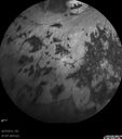
Retinitis Pigmentosa - Autosomal Dominant - Vision Loss at Young Age - Infrared598 views 54-year-old woman has autosomal dominant retinitis pigmentosa. Her mother and grandmother both had it and she has one son who has it and one daughter who doesn’t. She also has a sister with it. She lost vision since she was younger. She had cataract surgery done in January and February of this year. She has been seeing unusual flashing lights throughout the periphery and central vision for about the last year or two.
VISUAL ACUITY: OD 20/160, OS 20/160
|
|

Primary Ocular Lymphoma - 051812 598 views
|
|

AVM598 viewsFA of AVM in OS of a 48 yr old female.
|
|

Self Sealed Retinal Arteriol Macroaneurysm - one month after presentation - edema nearly gone598 viewsSD OCT line scan
|
|

Cystoid macular edema 4 months following cataract surgery with serous retinal detachment598 views68 year old with 2 months of vision loss OS 20/100. ECCE was 4 months ago.
|
|

Acute Posterior Multifocal Placoid Pigment Epitheliopathy - 4 years after onset598 views4 years after presentation - vision 20/80 OD 20/16 OS and there is subretinal fibrosis
|
|

Orbital Cellulitis - Optic nerve compression with ocular ischemia598 views81 year old woman with constricted eye movement, orbital cellulitis and severe vision loss in the right eye (LP). She has whitening of the retina and the OCT shows ischemic changes as well as subretinal fluid and blood. The vision in this eye declined to NLP and the eye remained ischemic despite control of her infection.
|
|

Myelinated Nerve Fiber Layer - Optic Nerve598 viewsRight eye subtle and inferior - left eye obvious
|
|

Central Retinal Vein Occlusion with Macular Edema598 views74 year old female The vision in the left eye has gotten worse over the last 3 weeks.
VA OD: Dcc20/25
VA OS: DccCF 3ft
IOP: TP: OD:16 OS:14
Vision improved to 20/40 in 3 months with monthly Aflibercept
|
|

Perforating Needle injury retrobulbar anesthetic - excellent final vision598 views71 year old man who had a vitrectomy for macular pucker and then developed a retinal detachment. There was an accidental needle stick during a retrobulbar anesthetic. The posterior puncture site is just inferior to the macula. The patient had a vitrectomy the next day for the retinal detachment and then 3 months later a vitrectomy to remove the fibrous proliferation from the puncture site. The final visual acuity was 20/20.
|
|

Serpiginous choroiditis597 viewsInactive scars in both eyes
|
|
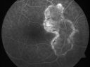
Serpiginous choroiditis597 viewsInactive scars in both eyes
|
|

Cytomegalovirus Retinitis - Partially Treated and AIDS retinopathy597 views44-year-old man a month agoe was diagnosed with HIV and he had pneumonia. He has had blurred vision for about a month, more in the left eye than the right eye. He has not yet seen anyone to start anti-AIDS therapy. He was given a medical doctor's name for a follow-up visit. He thinks his CD4 count is somewhere in the low 100s. Vision OU is 20/30
|
|
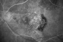
Wet Macular Degeneration Left Eye with Gutter (atypical) and Geographic Atrophy Right Eye597 views80-year-old man has wet age-related macular degeneration in his left eye. He has an atypical angiogram and that there is a gutter under the choroidal neovascular membrane suggesting still leakage of subretinal fluid. OS 20/400.
|
|

Chronic Central Serous Retinopathy treated with Photodynamic Laser and Visudyne597 views48-year-old man has had waxing and waning vision in the left eye for the last year from central serous retinopathy. OD is 20/16, OS is 20/30.
|
|

Wet Macular Degeneration with Juxtapapillary Neovascular Membrane and Choroidal Laser Treatment597 views87-year-old woman has wet age-related macular degeneration in the left eye with an extrafoveal choroidal neovascular membrane OD is 20/25, OS is 20/25
|
|

Pistol to head in attempted suicide 3 Months Ago - Retinal Atrophy - Shockwave Retinopathy VA Right Eye - Light Perception - Left Eye - 20/400597 views66-year-old woman had a gunshot wound to the head on August 12, 2010, with the entry wound just on the right side, just about below her temple, toward the lower part of the temple and the exit wound on the left side just above her cheek, a little behind the eye. Since then she recovered and did not need any surgery. When that all cleared she realized her vision was poor in both eyes. She has seen a neuro-ophthalmologist who thought she had traumatic optic neuropathy. OD light perception, OS 20/400.
|
|

Plaquenil (Hydroxycholorquine) Toxicity - Bull's Eye Maculopathy - 73 year old womand 25 Years therapy - 64 inches tall - 110 lbs597 views73-year-old woman has been on Plaquenil for twenty five years. She noticed loss of paracentral vision a few months ago. VISUAL ACUITY: OD 20/70, OS 20/30. Her color vision was 0 out 10 correct in both eyes and she also missed the controls.
|
|

Retinitis Pigmentosa Sine Pigmento or Unknown Dystrophy - Peripheral Superficial White Spots and Recurrent CME - FA MID597 views46-year-old woman three of her 5 siblings have been diagnosed with retinitis pigmentosa. Her mother and father were seen and as far as everyone can tell, neither of them has it, although there was some thought they might be carriers. They were evaluated twenty five years ago and since then as far as she knows, there has not been a lot of testing done.
She has noticed a substantial decline in vision in both eyes over the last month or two. OD 20/40, OS 20/50. IOP: Both eyes have CME
|
|

Gonioscopy, Blood in the Anterior Chamber from Hyphema597 viewsPatient comes in with blunt trauma to the right eye due to a BB gun incident. Patient was present with a hyphema at 8-o'clock about 1mm thick. Gonioscopy photos were then taken to show blood from the hyphema entered into the anterior chamber. Patient had no angle recession in the right eye.
|
|

Acute Multifocal Vitelliform Exudative Maculopathy Left Eye At 3 Mo Follow Up597 views
|
|

Central Areolar Choroidal Dystrophy597 viewsPatient wanted second opinion for atrophic macular degeneration. VA is 20/400, right eye and 20/100. left eye. Patient has very poor vision and also hearing impaired. Fundiscopic exam reveals very atrophy in the macula. FAF shows a central hole to the choroid with no neovascularization present.
|
|

Fibrosis Traction597 viewsYoung female patient with severe diabetes. Patients VA is 20/400 in the right eye. Fundus photos show a lot of fibrosis traction throughout the macula area. Fovea seems flat. Patient will undergo surgery for peeling of traction.
|
|

Proliferative Diabetic Retinopathy - Moderate - mild NVD with NVE 597 viewsFA shows leakage from neovascularization and dark areas in periphery from non-perfusion
|
|
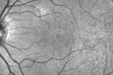
Outer Retinal Holes - Probably Solar Retinopathy (history is negative)597 views
|
|

Choroideremia - Female Carrier - Patchy Atrophy of Choroid and RPE - Lyonization597 views
|
|

Presumed Ocular Histoplasmosis with new CNVM in right and better eye - VA 20/80 OD; 20/800 OS597 viewsFluorescein Angiogram
|
|

Acute Posterior Multifocal Placoid Pigment Epitheliopathy - 4 years after onset597 views4 years after presentation - vision 20/80 OD 20/16 OS and there is subretinal fibrosis
|
|

Bilateral Branch Retinal Artery Occlusion - Intraarterial (hollenhorst) plaques visible597 views67 year old with acute visual field loss in each eye. Vision is 20/25 right eye 20/32 left eye. Images show intra-arterial plaques in each eye. The right eye on the optic nerve and the left eye in a branch retinal artery.
|
|

PDR NVD left eye and NVE right eye597 views41 year old diabetic woman with PDR in both eyes. High risk in the left eye. She had PRP, Avastin, and vitrectomy x 2 in the left eye with final vision of 20/25. The right eye had eventually PRP laser
|
|

Chronic Central Serous Chorioretinopathy Both Eyes597 views60 year old man with 20 year history of chronic CSR. He has had laser and PDT in the past. The left eye has been 20/100 for decades. The right eye is now declining. Was 20/32 and is down to 20/50. There is chronic low grade subretinal fluid away from the center in the right eye. (focal laser in the right eye in 2005, left eye 2001 at BPEI). Refampin tried in 2012, Aldactone in 2013. PDT in 204 and 2017 Images are from 2017
|
|

Chronic Central Serous Chorioretinopathy Both Eyes597 views60 year old man with 20 year history of chronic CSR. He has had laser and PDT in the past. The left eye has been 20/100 for decades. The right eye is now declining. Was 20/32 and is down to 20/50. There is chronic low grade subretinal fluid away from the center in the right eye. (focal laser in the right eye in 2005, left eye 2001 at BPEI). Refampin tried in 2012, Aldactone in 2013. PDT in 204 and 2017 Images are from 2017
|
|

Choroidal Nevus - Macula - Flat597 views37 year old female with 5 year stable nevus Left eye - OCT shows flat nevus (EDI scan)
|
|

Severe PDR with preretinal fibrosis597 views71 year old female untreated for a year following injections because of hurricane Irma. VA 20/30 OD and 5/200 OS. Sudden vision loss 1 month ago left eye was likely an ischemic event. Right eye is being treated with injections and PRP laser.
|
|

Peripheral exudative hemorrhagic chorioretinopathy - PECHR - Subfoveal fluid597 views85 year old female with
Fluctuating vision in the right eye.
VA 20/200 OD, 20/32 OS
|
|

Macular Hole Closed with Vitrectomy - Post-op 20/60 Vision596 views
|
|

26-year-old man choroideremia OU is 20/60596 views26-year-old man choroideremia OU is 20/60
|
|

Group 2a Juxtafoveal Retinal Telangiectasis (type 2)596 views58-year-old man decreased vision right eye and not better since cataract surgery one year ago. OD 20/40, OS 20/40.
|
|

Central Retinal Vein Occlusion - New- and old Macular Scar from AMD596 views87-year-old woman with longstaning macular scars and new vision loss left eye for 2 months. OD: 20/200, OS 20/400.
|
|

Angioid Streaks - New Extrafoveal Choroidal Neovascular Membrane Right Eye596 views61-year-old woman twelve days ago, saw a spot to the right of her central vision. OD is 20/25, OS is 20/25
Treated with one year of avastin with good result and no vision loss.
|
|

Acute Central Serous Chorioretinopathy with Symptoms in Hazy Vision Both Eyes596 views48-year-old man with hazy vision in the right eye for about three days. He has a little more of a headache than usual. He is under a fair amount of stress. He does not take any steroids that he knows of. He does use a nasal spray, which he is not sure if that is a steroid spray or not. He is going to call us with that medicine. I checked his blood pressure today is was 115/95.
VISUAL ACUITY: OD 20/15, OS 20/25
|
|
| 18117 files on 182 page(s) |
 |
|
|
|
|
50 | |
|
|
|
|


