|
|

Retinitis Pigmentosa Sine Pigmento or Unknown Dystrophy - Peripheral Superficial White Spots and Recurrent CME596 x angesehen46-year-old woman three of her 5 siblings have been diagnosed with retinitis pigmentosa. Her mother and father were seen and as far as everyone can tell, neither of them has it, although there was some thought they might be carriers. They were evaluated twenty five years ago and since then as far as she knows, there has not been a lot of testing done.
She has noticed a substantial decline in vision in both eyes over the last month or two. OD 20/40, OS 20/50. IOP: Both eyes have CME
|
|

Retinitis Pigmentosa Sine Pigmento or Unknown Dystrophy - Peripheral Superficial White Spots and Recurrent CME - FA MID596 x angesehen46-year-old woman three of her 5 siblings have been diagnosed with retinitis pigmentosa. Her mother and father were seen and as far as everyone can tell, neither of them has it, although there was some thought they might be carriers. They were evaluated twenty five years ago and since then as far as she knows, there has not been a lot of testing done.
She has noticed a substantial decline in vision in both eyes over the last month or two. OD 20/40, OS 20/50. IOP: Both eyes have CME
|
|

Non-specific (uncharaterized / unknown) Macular Dystrophy - FAF596 x angesehen59-year-old man has macular dystrophy in both eyes. He had some vision changes in medical school in the 1980s and saw Dr. Gass for that. He had some pigment epithelial irregularities in both eyes. He had strabismus as a child and had muscle surgery. He is color blind, along with many people in his family, and as far as he knows, it is congenital.
20/25, 20/30
|
|

Fibrosis Traction596 x angesehenYoung female patient with severe diabetes. Patients VA is 20/400 in the right eye. Fundus photos show a lot of fibrosis traction throughout the macula area. Fovea seems flat. Patient will undergo surgery for peeling of traction.
|
|

Papilledema - Sagittal Cerebral Vein Thrombosis - Lung Transplant Cystic Fibrosis 36 Year Old Man596 x angesehenLine scan through optic nerve
|
|

Vitreomacular traction - recent vision loss left eye - FA596 x angesehen
|
|

AVM596 x angesehenFA of AVM in OS of a 48 yr old female.
|
|

Bilateral Macular Star596 x angesehenYoung female patient comes in with blurred vision in both eyes. VA is 20/40 in both eyes. Fundus photos show visible macular star centrally in both eyes. This is a result of Bilateral Neuroretinitis due to cat scratch.
|
|

Iris Nevus596 x angesehenPatient came in for cataract evaluation. VA 20/200, right eye, 20/60, left eye. Slit lamp photography shows iris nevus 3-4 mm located nasally at 9-o'clock in the left eye.
|
|

Acute Macular Neuroretinopathy - (AMN) - Spot in Vision OS - 40 Year old Female596 x angesehenFluorescein Angiogram is normal
|
|

Choroideremia - Female Carrier - Patchy Atrophy of Choroid and RPE - Lyonization596 x angesehen
|
|

Posterior Uveal EffusionPosterior Uveal Effusion596 x angesehen
|
|

Presumed Ocular Histoplasmosis with new CNVM in right and better eye - VA 20/80 OD; 20/800 OS596 x angesehenFluorescein Angiogram
|
|

Choroidal Folds Hyperope (+5.00)596 x angesehen
|
|

Trousseau Sign - CRVO and Pancreatic Cancer596 x angesehenLeft eye shows CRVO with severe edema. The right eye has multiple small hemorrhages. Some of this is probably anemic retinopathy and some from hypercoaguability of cancer. The patient has pancreatic cancer.
|
|

Orbital Cellulitis - Optic nerve compression with ocular ischemia596 x angesehen81 year old woman with constricted eye movement, orbital cellulitis and severe vision loss in the right eye (LP). She has whitening of the retina and the OCT shows ischemic changes as well as subretinal fluid and blood. The vision in this eye declined to NLP and the eye remained ischemic despite control of her infection.
|
|

Bullous Retinoschisis - Confirmed with SD OCT596 x angesehenAtypical location for retinoschisis - SD-OCT is used to confirm that this is retinoschisis and not a retinal detachment
|
|

Syphilitic Chorioretinitis596 x angesehenSevere outer retinal atrophy and RPE non-geographic atrophy from syphilis. Vision improved from 20/63 to 20/32 with therapy. Images look a lot like DUSN. Patient had several surgeries (vitrectomy for floaters, vitrectomy for macular pucker, Avastin injections and macular laser for edema) prior by other retina specialists prior to being correctly diagnosed and treated.
|
|
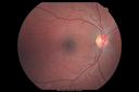
Torpedo Maculopathy596 x angesehenLikely a variant of CHRPE
|
|

Chronic Central Serous Chorioretinopathy Both Eyes596 x angesehen60 year old man with 20 year history of chronic CSR. He has had laser and PDT in the past. The left eye has been 20/100 for decades. The right eye is now declining. Was 20/32 and is down to 20/50. There is chronic low grade subretinal fluid away from the center in the right eye. (focal laser in the right eye in 2005, left eye 2001 at BPEI). Refampin tried in 2012, Aldactone in 2013. PDT in 204 and 2017 Images are from 2017
|
|

Choroidal Nevus - Macula - Flat596 x angesehen37 year old female with 5 year stable nevus Left eye - OCT shows flat nevus (EDI scan)
|
|

Severe PDR with preretinal fibrosis596 x angesehen71 year old female untreated for a year following injections because of hurricane Irma. VA 20/30 OD and 5/200 OS. Sudden vision loss 1 month ago left eye was likely an ischemic event. Right eye is being treated with injections and PRP laser.
|
|

Wet AMD 62 year old male smoker 595 x angesehen62-year-old man has wet age-related macular degeneration in the right eye. He has been undergoing treatment with Lucentis for that. Then his left eye developed hazy vision a few weeks ago while he was overseas and now his vision is hazy in both eyes. OD 20/40, OS 20/30. FA shows a classic subfoveal choroidal neovascular membrane in the left eye, which is leaking and in the right eye, which is dry. He is going to stop smoking today. FU: VA 20/30 OU with 2 years of every 6 weeks bilateral avastin!
|
|

Diabetic Patient with Macular Edema and Blood Pressure 200/95595 x angesehen49-year-old decreasing vision over the last year. OD is 20/80, OS 20/80. blood pressure which was 200/95.
|
|

Serpiginous choroiditis595 x angesehenInactive scars in both eyes
|
|
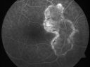
Serpiginous choroiditis595 x angesehenInactive scars in both eyes
|
|

Chronic Pseudophakic Cystoid Macular Edema in a Patient with Diabetes595 x angesehen74-year-old woman has chronic cystoid macular edema in the left eye. She has had vision loss intermittently in that eye for 2 years, the right eye, unfortunately, lost vision from a central retinal vein occlusion in 6 years ago.
|
|

Central Retinal Artery Occlusion Left Eye 2 Weeks Old - Still Non-Perfusion595 x angesehen85-year-old woman who had sudden severevision loss in the left eye two weeks ago. she can see a little more centrally and much more in the periphery, since about the last four days.
VISUAL ACUITY: OD 20/30, OS: Hand motion
|
|

595 x angesehen
|
|

Macular Pucker 20/40 vision and recent cataract surgery595 x angesehen 73-year-old man distortion and vision loss since ECCE 6 months ago OS. Patient taking anti-inflammatory drops. VISUAL ACUITY: OD 20/30, OS 20/40.
|
|

Macular Pucker - Diabetic - Severe non-foveal Edema OS595 x angesehen74-year-old man has a macular pucker in the left eye and diabetic retinopathy.
VISUAL ACUITY: Vision OD is 20/25, OS is 20/40.
|
|

Macular Pucker - 3 Weeks Vision Loss - 20/50 Vision - Preoperative Images595 x angesehenPREOP:
59-year-old man new onset floaters about a year ago. Subsequent to that his vision was not quite right in the right eye and then he noticed, starting about three weeks ago, substantial change in the vision. VISUAL ACUITY: Vision OD is 20/50, OS is 20/16
|
|
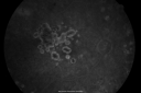
Diabetic Retinopathy - Foveal Laser Scar Right Eye595 x angesehen74-year-old woman has diabetic retinopathy in both eyes. She has had focal laser in her right eye done elsewhere in the mid 90’s and unfortunately the laser was fairly close to the center and as time progressed she developed foveal atrophy around the scars. She has noticed her vision declining since she was here last a year ago. She does take Travatan in both eyes at night for glaucoma.
VISUAL ACUITY: OD 20/200, OS 20/40.
|
|

Myopic Traction Maculopathy - Vitreoretinal Traction - Possible Occult Neovascular Membrane595 x angesehen58-year-old woman was seen in the office on June 30, 2011. In 2002, her vision was 20/30. Subsequent to that she had her left eye made her distance eye, which was dominate. She had LASIK surgery on the right eye to make that her near eye and she was getting along well until about a year ago when she noticed substantial vision loss in the left eye only. VISUAL ACUITY: The near eye is 20/200, but she reads J2. OS 20/100- and J16.
|
|

Acute Non-arteritic Anterior Ischemic Optic Neuropathy Right Eye - Previous NA-AION left eye 595 x angesehen69-year-old man previously lost vision in the left eye from nonarteritic anterior ischemic optic neuropathy in 2006. About a week ago and noticed blurred vision in the inferonasal part of the vision in the right eye.
VISUAL ACUITY: Vision OD is 20/16, OS is 20/40
|
|

Angioid Streaks and Macular Degeneration - CNVM OS - Band Keratopathy 595 x angesehen79-year-old man has wet age-related macular degeneration in the left eye, for which he is having Lucentis treatment. OD 20/40, Pinhole 20/32. OS 20/160, Pinhole 20/50. He has band keratopathy sparing the visual axis in both eyes. ICG shows angioid streaks both eye.
|
|

Retinitis Pigmentosa Sine Pigmento or Unknown Dystrophy - Peripheral Superficial White Spots and Recurrent CME595 x angesehen1 month of treatment with diamox 500 mg BID- cme is a little better
|
|
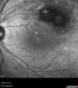
Retinal Arterial Macroaneurysm - Untreated Hypertension (250/135) - Serous Macular Detachment595 x angesehen82 year old asian woman. VA 20/80 Headaches and vision loss for 1 month. Negative medical history but has not been to the doctor for years. (no insurance)
|
|

Gonioscopy of Blood in the Angle595 x angesehenYoung female who has a history of uveitis, glaucoma and juvenile rheumatoid arthritis. VA is hand motion in the left eye. Pigment changes with some blood in the anterior chamber seen here on Gonioscopy.
|
|

Optic Disk Drusen - Pucker - Pattern Dystrophy595 x angesehenAutofluorescence from Drusen and Macular Dystrophy
|
|

Acute AION OS - Mild disc pallor OD595 x angesehenSudden vision loss OS x 1 week with optic nerve swelling. Evidence of optic atrophy in right eye.
|
|

Trousseau Sign - CRVO and Pancreatic Cancer595 x angesehenLeft eye shows CRVO with severe edema. The right eye has multiple small hemorrhages. Some of this is probably anemic retinopathy and some from hypercoaguability of cancer. The patient has pancreatic cancer.
|
|

Bilateral Branch Retinal Artery Occlusion - Intraarterial (hollenhorst) plaques visible595 x angesehen67 year old with acute visual field loss in each eye. Vision is 20/25 right eye 20/32 left eye. Images show intra-arterial plaques in each eye. The right eye on the optic nerve and the left eye in a branch retinal artery.
|
|

Wagner Syndrome 595 x angesehen33 year old man with family history of Wagner syndrome - the left eye had RD repair. The right eye has posterior peripheral pigmented lesions captured here superiorly
|
|

Torpedo Maculopathy595 x angesehenLikely a variant of CHRPE
|
|

Photic Maculopathy Following Cataract Surgery595 x angesehenInitial images are from 1 week and subsequent images from 1 month post surgery.
|
|

Multifocal Vitelliform Macular Dystrophy595 x angesehen58 year old female with mild vision loss - 20/20 OU - extensive testing has shown no cancer. Case of Karina Findlay, MD
|
|

Melanosis Oculi and Choroidal Melanoma595 x angesehen88 year old female with 2.5 mm elevated choroidal melanoma and melanosis oculi. She also has geographic atrophy from AMD. The right eye is the better eye with 20/40 vision. After observation with growth, brachytherapy was done.
|
|

Diabetic Macular Edema with Circinate Exudate Red Free594 x angesehen74-year-old man has diabetic macular edema in both eyes and background diabetic retinopathy. His vision has blurred over the last few months. His blood sugars have been fluctuating.
VISUAL ACUITY: Vision OD is 20/25, OS is 20/80
|
|
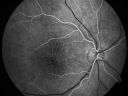
Pigmented Maculae - Mother and Daughter - Normal Visual Acuity594 x angesehen12-year-old had a dilated macular exam and the macula look abnormal. Interestingly, her mother, who is with her at this visit, was told about a year ago that her maculae are abnormal too. Her mother’s vision is fine. In the family tree, there are no known retinal dystrophies. They are both healthy.
VISUAL ACUITY: Her vision is 20/20 in both eyes.
|
|

Concentric Geograophic Atrophy - Age-related Macular Degeneration - Charles Bonnet Syndrome - Bulls Eye594 x angesehen88-year-old woman has age-related with vision gradually declining over the last year. She sees spots in her vision, like red spots and dark spots and she even notices during the daytime, for example when she was looking at her minister, it looked like there were little flowering pots coming out of his head.
VISUAL ACUITY: OD 20/200, OS 20/400.
|
|

Hemicentral Retinal Vein Occlusion Vision 20/60 594 x angesehen84-year-old man has a hemicentral retinal vein occlusion in the right eye. His vision has declined since then.
VISUAL ACUITY: Vision OD is 20/60.
|
|

AIDS - HIV Retinopathy - Cotton Wool Spots594 x angesehen54-year-old woman HIV positive for 18 years. Her CD4 count is currently less than 50 and she was hospitalized for pneumocystis carinii pneumonia 5 months ago. She has noticed just for the last few weeks a few blind spots in the paracentral vision. Predominantly out of her right eye when she closes her left eye, she sees a few temporal blind spots. VA 20/25 in both eyes.
|
|

Diabetic Macular Edema594 x angesehen63 yr old Male
VA 20/20 OD 20/25 OS
No complaints
|
|

Retinitis Pigmentosa Newly Diagnosed 55 Year Old594 x angesehen55-year-old noticed his vision declining recently, over the last few weeks, particularly in the right eye. He said previous to that the vision was not normal but was pretty good. OD is 20/100, OS is 20/50
OCT SCAN: There is macular thickening in both eyes with intraretinal cysts.
|
|

594 x angesehen63-year-old man diabetic for twenty two years. His central vision is fine.
VISUAL ACUITY: OD 20/25, OS 20/25
|
|

Serous Retinal Detachment - Hypertensive Retinopathy - 165/120 594 x angesehen48-year-old woman noticed when she was blinking there was a white flash in the left eye starting about a month ago. She sees it temporally. OD 20/16, OS 20/20. There is a serous retinal detachment superotemporal to the optic nerve falling short of the fovea. Her blood pressure 165/120.
|
|

Retinitis Pigmentosa Sine Pigmento or Unknown Dystrophy - Peripheral Superficial White Spots and Recurrent CME594 x angesehen46-year-old woman three of her 5 siblings have been diagnosed with retinitis pigmentosa. Her mother and father were seen and as far as everyone can tell, neither of them has it, although there was some thought they might be carriers. They were evaluated twenty five years ago and since then as far as she knows, there has not been a lot of testing done.
She has noticed a substantial decline in vision in both eyes over the last month or two. OD 20/40, OS 20/50. IOP: Both eyes have CME
|
|

Optic Nerve Hypoplasia - 43 Year Old Man - declining vision594 x angesehen43-year-old man was diagnosed with optic nerve hypoplasia at age fifteen at the National Institute of Health. His vision seems to be declining lately.
Vision OD is 20/80, OS is 20/120
|
|
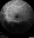
Uveitis - Retinitis - Vasculitis - possibly Syphilitis594 x angesehenVisit 1: 12-06-12
74-year-old man 12-3-12 visit. 2 months ago noticed vision loss, first in the right eye than the left. He is getting tired easily. He can’t walk for a long distance. He has lost five pounds within the last month. He also had a severe itchy episode, but not quite a rash a few days ago, which went away after he took a shower. FTA-ABS is positive but a spinal tap, which was negative. OD 1/200, OS 20/63.
|
|

Vasculitis - Retinitis - Uveitis - Vision NLP right eye , 20/50 left eye594 x angesehen
|
|

Asymptomatic Juxtapapillary Choroidal Neovascular Membrane - Reticular Macular Disease (Wet AMD) 594 x angesehen
|
|

FAF594 x angesehen
|
|

Serous Macular Detachment Both Eyes - Wet AMD vs. CSR - 62 Year Old Man - Color Photo594 x angesehen
|
|

Acute Multifocal Vitelliform Exudative Maculopathy Left Eye At 3 Mo Follow Up594 x angesehen
|
|

Proliferative Diabetic Retinopathy and Vitreous Hemorrhage - FA shows leakage of dye from NVE594 x angesehen
|
|

Vitreomacular traction - recent vision loss left eye - FA594 x angesehen
|
|

Branch Retinal Vein Occlusion with worse macular edema from hypertension - Photo from 2008 with initial occlusion - Vision 20/40594 x angesehen
|
|

Retinal Artery Occlusion594 x angesehenPatient comes in for eval on artery occlusions in both eyes. VA is 20/400, right eye and NLP, left eye. Fundus photos show paniretinal scars in the right eye with arterial narrowing and the left eye has arterial narrowing as well.
|
|

Acute Macular Neuroretinopathy - (AMN) - Spot in Vision OS - 40 Year old Female594 x angesehenFluorescein Angiogram is normal
|
|
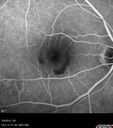
Pre-retinal hemorrhage following Posterior Vitreous Detachment (separation - PVD)594 x angesehen68 year old woman with acute PVD OD and new preretinal hemorrhage - also there is a macular nevus
|
|

Late Onset Retinal Degeneration (L-ORD)594 x angesehen55 year old with acute vision loss from a CNVM in the right eye. He responded to Lucentis therapy. His mother and her family has been confirmed genetically to have L-ORD and were part of the early reports.
|
|

Subretinal Hemorrhage - Commotio Retinae - Macula - hit with baseball594 x angesehen15 year old with VA 20/60 3 days after being hit with baseball in the right eye
|
|
594 x angesehen
|
|

Pre retinal hemorrhage associated with partial PVD594 x angesehen56 year old female in for checkup- no symptoms and normal vision - there is a hemorrhage by the nerve that could be vascular, tractional or from her mild glaucoma (which is controlled). The macular hemorrhage is preretinal.
|
|

Macular Hole Closed with Vitrectomy - Preop593 x angesehen
|
|

Acute Central Serous Chorioretinopathy with Symptoms in Hazy Vision Both Eyes593 x angesehen48-year-old man with hazy vision in the right eye for about three days. He has a little more of a headache than usual. He is under a fair amount of stress. He does not take any steroids that he knows of. He does use a nasal spray, which he is not sure if that is a steroid spray or not. He is going to call us with that medicine. I checked his blood pressure today is was 115/95.
VISUAL ACUITY: OD 20/15, OS 20/25
|
|
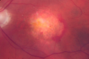
Wet Macular Degeneration Left Eye with Gutter (atypical) and Geographic Atrophy Right Eye593 x angesehen80-year-old man has wet age-related macular degeneration in his left eye. He has an atypical angiogram and that there is a gutter under the choroidal neovascular membrane suggesting still leakage of subretinal fluid. OS 20/400.
|
|

80 Year old man with 3 day history of vision loss right eye. Vision 4/200.593 x angesehen
|
|
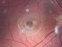
Macular Hemorrahge and Probable CNVM Right eye for 2 weeks VA 20/400 - 20 Year old593 x angesehen20-year-old man noticed vision loss in the right eye about two weeks ago and if anything it seems to be getting worse. He is a high myope. VISUAL ACUITY: OD 20/400
|
|

Diabetic Macular Edema and Hypertensive Retinopathy - Circinate Exudate (Ring Exudate) 593 x angesehen55-year-old woman diabetic for fifteen years and high blood pressure, especially over the last few months. She has had problems with nosebleeds, headaches, and there has been some difficulty bringing her blood pressure down. She said now the blood pressure is under control but it was running 220/120 mmHg for some time. Her vision in the right eye has been poor for four weeks with a spot in the central vision and both eyes have been blurred.
VISUAL ACUITY: Vision OD is 20/160, OS is 20/60
|
|

Macular Pucker 20/30 Good Vision 593 x angesehen64-year-old woman decreased vision in the left eye for about six months. You saw a wrinkle on the eye and suggested she come in here for an evaluation. She had radial keratotomy done 20 years ago.
VISUAL ACUITY: OD 20/25, OS 20/30
|
|

Non-Ischemic Central Retinal Vein Occlusion in Young Man (30 years) - Many Cotton Wool Spots593 x angesehen30-year-old man with a history of a central retinal vein occlusion for which he was first examined on 2 months ago and his vision then was 4/200. He responded nicely to two Avastin treatments. OD is 20/20, OS is 20/60
|
|

Chronic Pseudophakic Cystoid Macular Edema - Haptic Migrated into Anterior Chamber Through Peripheral Iridotomy593 x angesehen57-year-old man with congenital nystagmus. He had cataract surgery done 30 years ago and has decreased vision left eye for 6 months. OD is 20/50, OS is 20/100
|
|

Choroidal Melanoma (Medium) Exudative Retinal Detachment 593 x angesehen87-year-old man noticed a new onset floater a few months ago. OD is 20/40, OS is 20/30
|
|

Punctate Inner Choroidopathy - Recurrent CNVM 4 years after initial treatment Left Eye - Fluorescein Angiogram593 x angesehenJune 7, 2012. This pleasant 35-year-old woman noticed decreasing vision in the left eye recently. She returns now noticing the vision in the left eye is declining. OD 20/50, OS 20/80. OS: Vertical C/D ratio is 0.3. There is a scar just inferonasal to the fovea, which has grown some towards the fovea with a little blood just under the fovea.
|
|
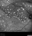
Primary Ocular Lymphoma - 020513 593 x angesehen
|
|

BRVO Inferiorly with Retinal Hemorrhages in the Left Eye593 x angesehenPatient comes in with complaint of "spot in vision", sort of a cloudy haze in the left eye. Patient's VA was 20/100-1 with no improvement with pinhole in the left eye. Fundus photos show multiple retinal hemorrhages scattered with a branch retinal vein occlusion inferiorily. Patient is also diabetic.
|
|

BDR OD with Macular Edema593 x angesehen
|
|

Pre-proliferative Diabetic Retinopathy - Peripheral non-perfusion visible on FA593 x angesehen
|
|

Retinal Arterial Macroaneurysm - Untreated Hypertension (250/135) - Serous Macular Detachment593 x angesehen82 year old asian woman. VA 20/80 Headaches and vision loss for 1 month. Negative medical history but has not been to the doctor for years. (no insurance)
|
|

Proliferative Diabetic Retinopathy - Moderate - mild NVD with NVE 593 x angesehenFA shows leakage from neovascularization and dark areas in periphery from non-perfusion
|
|

593 x angesehen
|
|

Exogenous Fungal Endophthalmitis593 x angesehen69 year old female with endophthalmitis 2 months after ECCE. Treated with vitrectomy and 3 injections of amphotericin B. Vision improved from 5/200 - 20/50
|
|

Late Onset Cone Dystrophy - Very Mild593 x angesehenOnly IR and OCT show very mild bull's eye. 67 year old female with difficulty seeing in bright light
|
|

Occult Macular Dystrophy - 9 Years later 593 x angesehen53 year old man. Vision is better at 20/50 OU
|
|

Central Retinal Vein Occlusion with Macular Edema593 x angesehen74 year old female The vision in the left eye has gotten worse over the last 3 weeks.
VA OD: Dcc20/25
VA OS: DccCF 3ft
IOP: TP: OD:16 OS:14
Vision improved to 20/40 in 3 months with monthly Aflibercept
|
|
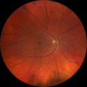
Heterochromia - Left Fundus - Possible Waardenburg Syndrome593 x angesehen49 year old female with normal vision. No hearing loss. Fundus in the left eye has large patchy hypopigmentation. OCT in that area shows decrease vascularity.
|
|

Peripheral exudative hemorrhagic chorioretinopathy - PECHR - Subfoveal fluid593 x angesehen85 year old female with
Fluctuating vision in the right eye.
VA 20/200 OD, 20/32 OS
|
|
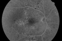
BDR and Diabetic Macular Edema with Fresh Laser Right Eye592 x angesehen60-year old man OD 20/60, OS 20/50 worsening vision both eyes
|
|
| 18117 Dateien auf 182 Seite(n) |
 |
|
|
|
|
51 | |
|
|
|
|


