|
|

Polypoidal Choroidal Vasculopathy with Pulsating ICG polyp left eye594 views
|
|

Acute Macular Neuroretinopathy - (AMN) - Spot in Vision OS - 40 Year old Female594 viewsFluorescein Angiogram is normal
|
|

Choroidal Effusions and Serous retinal detachment with Systemic Testosterone and Cialis in 75 year old Man594 viewsFluorescein angiogram shows some staining of the disk - confocal system renders peripheral effusions black
|
|

594 views
|
|

West Nile Virus Multifocal Choroiditis and diabetic retinopathy594 viewsMultifocal and clustered focal lesions are from west nile virus. The retinopathy is probably diabetic but could also be related to west nile virus
|
|

Occult Macular Dystrophy - 9 Years later 594 views53 year old man. Vision is better at 20/50 OU
|
|
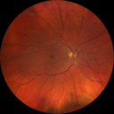
Heterochromia - Left Fundus - Possible Waardenburg Syndrome594 views49 year old female with normal vision. No hearing loss. Fundus in the left eye has large patchy hypopigmentation. OCT in that area shows decrease vascularity.
|
|

Peripheral exudative hemorrhagic chorioretinopathy - PECHR - Subfoveal fluid594 views85 year old female with
Fluctuating vision in the right eye.
VA 20/200 OD, 20/32 OS
|
|
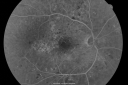
BDR and Diabetic Macular Edema with Fresh Laser Right Eye593 views60-year old man OD 20/60, OS 20/50 worsening vision both eyes
|
|

Serpiginous choroiditis593 viewsInactive scars in both eyes
|
|

Acute Central Serous Chorioretinopathy with Symptoms in Hazy Vision Both Eyes593 views48-year-old man with hazy vision in the right eye for about three days. He has a little more of a headache than usual. He is under a fair amount of stress. He does not take any steroids that he knows of. He does use a nasal spray, which he is not sure if that is a steroid spray or not. He is going to call us with that medicine. I checked his blood pressure today is was 115/95.
VISUAL ACUITY: OD 20/15, OS 20/25
|
|

Wet Macular Degeneration Left Eye with Gutter (atypical) and Geographic Atrophy Right Eye593 views80-year-old man has wet age-related macular degeneration in his left eye. He has an atypical angiogram and that there is a gutter under the choroidal neovascular membrane suggesting still leakage of subretinal fluid. OS 20/400.
|
|

Ruptured Retinal Arterial Macroaneurysm - Submacular Hemorrhage - Vision 20/400 never recovered - Hypertension 205/94593 views
|
|

80 Year old man with 3 day history of vision loss right eye. Vision 4/200.593 views
|
|
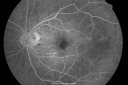
Ocular Ishemic Syndrome - Bilateral Carotid Occlusions - OD 20/60 OS 20/200 Enlarged Foveal Avascular Zone and Temporal Non-perfusion593 views75-year-old man had laryngeal cancer treated in 15 year ago with radiation. He recently was told that the carotid circulation is closed off on both sides. He does have a patent vertebral artery, which is supplying him pretty well and in addition to that his cardiac output is compromised by poor mitral valves. His consideration is to what can be done about that in the future.
VISUAL ACUITY: OD 20/60, OS 20/200
|
|

Chronic - Old Diabetic Tractional Retinal Detachment Left Eye593 views77-year-old woman Vision OD is 20/40, OS is 7/200OD: Vertical C/D ratio is 0.3. There is panretinal laser. The macula is flat and attached.
OS: Vertical C/D ratio is 0.3. There is panretinal laser. There is a tractional retinal detachment with preretinal fibrosis nasal to the optic nerves.
|
|

X-linked Juvenille Retinoschisis - Peripheral Retinal Vascular Anomalies - Vitreous Hemorrhage - 8 Year Old Boy593 views8-year-old child OD 20/40, OS 20/50
OD: Vertical C/D ratio is 0.2. There are foveal cysts. There is also a retinal elevation inferiorly and there are patchy peripheral retinal hemorrhages.
OS: Vertical C/D ratio is 0.2. There are foveal cysts. There is peripheral retinal issues with some hemorrhage in some areas of peripheral retinoschisis.
|
|

Cotton Wool Spot593 views
|
|

Punctate Inner Choroidopathy - Recurrent CNVM 4 years after initial treatment Left Eye - Fluorescein Angiogram593 viewsJune 7, 2012. This pleasant 35-year-old woman noticed decreasing vision in the left eye recently. She returns now noticing the vision in the left eye is declining. OD 20/50, OS 20/80. OS: Vertical C/D ratio is 0.3. There is a scar just inferonasal to the fovea, which has grown some towards the fovea with a little blood just under the fovea.
|
|
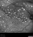
Primary Ocular Lymphoma - 020513 593 views
|
|

Asymptomatic Juxtapapillary Choroidal Neovascular Membrane - Reticular Macular Disease (Wet AMD) 593 views
|
|

Reticular Macular Disease (Pseudo-drusen) Both Eyes - Occult CNVM and Macular Edema Right Eye 593 viewsFluorescein Angiogram
|
|

Retinal Arterial Macroaneurysm - Untreated Hypertension (250/135) - Serous Macular Detachment593 views82 year old asian woman. VA 20/80 Headaches and vision loss for 1 month. Negative medical history but has not been to the doctor for years. (no insurance)
|
|

Retinitis Pigmentosa Sine Pigmento 593 views
|
|

Acute AION OS - Mild disc pallor OD593 viewsSudden vision loss OS x 1 week with optic nerve swelling. Evidence of optic atrophy in right eye.
|
|

CRAO WITH SPARING OF CILIORETINAL ARTERY593 viewsThis patient was found to have retinal edema from a Central Retinal Artery Occlusion with sparing of the cilioretinal artery. There were no discernible Hollenhorst plaques
|
|

Sickle Retinpoathy SC Disease - Vitreous Hemorrhage Left Eye - 29 Year old593 views
|
|

Posterior Uveal Effusion593 views
|
|

Presumed Ocular Histoplasmosis with new CNVM in right and better eye - VA 20/80 OD; 20/800 OS593 viewsFluorescein Angiogram
|
|

OHS CNVM post treatment with Lucentis - VA now 20/25593 views
|
|

Retinal Atrophy 1 year following Streptococcus sanguinis endophthalmitis - Vision 5/200593 views79 year old woman with sleep apnea who sleeps with CPAP machine
|
|

Branch Retinal Vein Occlusion (BRVO) no macular edema - cotton wool spot593 views85 year old man with asymptomatic BRVO - his vision stayed great without needing any treatment.
|
|

Branch Retinal Vein Occlusion (BRVO) no macular edema - cotton wool spot593 views85 year old man with asymptomatic BRVO - his vision stayed great without needing any treatment.
|
|

Torpedo Maculopathy593 viewsLikely a variant of CHRPE
|
|

Late Onset Cone Dystrophy - Very Mild593 viewsOnly IR and OCT show very mild bull's eye. 67 year old female with difficulty seeing in bright light
|
|

Central Retinal Vein Occlusion with Macular Edema593 views74 year old female The vision in the left eye has gotten worse over the last 3 weeks.
VA OD: Dcc20/25
VA OS: DccCF 3ft
IOP: TP: OD:16 OS:14
Vision improved to 20/40 in 3 months with monthly Aflibercept
|
|

Stargart's Macular Dystrophy Severe592 views32-year-old man carries a diagnosis of Stargardt’s disease with poor vision in both eyes since he was 13. He has not noticed any substantial change since he was here 4 years ago.
VISUAL ACUITY: OD 10/200, OS 8/200
|
|

Wet Macular Degeneration with Juxtapapillary Neovascular Membrane and Choroidal Laser Treatment592 views87-year-old woman has wet age-related macular degeneration in the left eye with an extrafoveal choroidal neovascular membrane OD is 20/25, OS is 20/25
|
|

80 Year old man with 3 day history of vision loss right eye. Vision 4/200.592 views
|
|

Macular Hole - Traumatic - 11 year old boy (also retinal whitening Berlins Edema)592 views11-year-old boy with a history of blunt trauma to the left eye yesterday. He reports he lost vision immediately but it has gotten somewhat better. He reports he sees “red streamers†and he has very blurry visual acuity., 20/200 OS
|
|

Hyperpigmentation of Retinal Pigment Epithlium in Dry AMD (hyperplasia)592 views87-year-old woman has wet age-related macular degeneration in the left eye. She had treatment up north but there has been a gap in therapy and the vision in the left eye is poor. Vision OD is 20/25, OS is 20/400
|
|

Myopic Traction Maculopathy - Vitreoretinal Traction - Possible Occult Neovascular Membrane592 views58-year-old woman was seen in the office on June 30, 2011. In 2002, her vision was 20/30. Subsequent to that she had her left eye made her distance eye, which was dominate. She had LASIK surgery on the right eye to make that her near eye and she was getting along well until about a year ago when she noticed substantial vision loss in the left eye only. VISUAL ACUITY: The near eye is 20/200, but she reads J2. OS 20/100- and J16.
|
|

Eccentric Disciform Scar - Breakthrough Vitreous Hemorrhage - InferoTemporal Subretinal Fibrosis - Ectopic Choroidal Neovascular Membranes592 views88-year-old man has noticed for the last few months the vision in the right eye is foggy, like there is a film in the eye or a haze, His vision is 20/16 in each eye.
Right eye - inferotemporal periphery there is a hemorrhage at the equator at about 7:00 o’clock, which is about 4-disc diameters across and adjacent to that there is scarring.
Left eye - large disciform scar inferotemporally
|
|

Macular and Peripheral Retinal Scarring from Diabetic Laser Done 25 years previously592 views72-year-old man had macular and peripheral laser done in the past(1980's)(25 years ago). His vision, although limited, seems about stable to him. He is diabetic for 30 years. OD is 20/160, OS is 20/160.
|
|

Congenital Achromatopsia 592 views26-year-old man was diagnosed with congenital achromatopsia by ERG as a child. He has family members who also have this disorder. OD 20/200, OS 20/200.
|
|

Choroidal Melanoma (Medium) Exudative Retinal Detachment 592 views87-year-old man noticed a new onset floater a few months ago. OD is 20/40, OS is 20/30
|
|

Macular Hole Closed - Nerve Fiber Layer Defects Visible after Internal Limiting Membrane Peel - Vision 20/60592 views53-year-old woman had vitrectomy for a stage IV macular hole in the left eye on June 1st. Her vision has improved some but still has a bump in it.
VISUAL ACUITY: Vision OS is 20/80, PH is 20/60
|
|
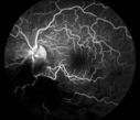
Cytomeglovirus592 views25 year old male experiencing blurry vision OS for two days. Onset very rapid. VA 20/40 OS 20/30 OD. After HIV titer test, determined the pt. had HIV and Cytomeglovirus.
|
|

Alphamethlyacyl COA Racemase Deficiency592 views
|
|

Punctate Inner Choroidopathy - Recurrent CNVM 4 years after initial treatment Left Eye 592 viewsJune 7, 2012. This pleasant 35-year-old woman noticed decreasing vision in the left eye recently. She returns now noticing the vision in the left eye is declining. OD 20/50, OS 20/80. OS: Vertical C/D ratio is 0.3. There is a scar just inferonasal to the fovea, which has grown some towards the fovea with a little blood just under the fovea.
|
|

Pigmented Peripheral Retinal Degeneration592 views42-year old male comes in for routine eye exam and to follow up on peripheral retinal degeneration in both eyes. VA is 20/20, right eye and 20/25, left eye. Patient is asymptomatic with no visual complaints.
|
|

Uveal Choroidal Melanoma592 viewsPatient comes in for evaluation on a Choroidal Melanoma in the right eye. VA was 20/25 in both eyes. The melanoma is in the temporal aspect of the right eye. It measured at 0.7mm elevated after doing a BSCAN Ultrasound.
|
|

Reticular Macular Disease (Pseudo-drusen) Both Eyes - Occult CNVM and Macular Edema Right Eye 592 viewsFluorescein Angiogram
|
|

Reticular Macular Disease (Pseudo-drusen) Both Eyes - Occult CNVM and Macular Edema Right Eye 592 viewsFluorescein Angiogram
|
|

Reticular Macular Disease (Dry Macular Degeneration) - Spectral Domain Optical Coherence Tomography Line Scan592 views85 year old woman with 20/25 vision OU and reticular macular disease
|
|

Proliferative Diabetic Retinopathy - Moderate - mild NVD with NVE 592 viewsFA shows leakage from neovascularization and dark areas in periphery from non-perfusion
|
|

Acute AION OS - Mild disc pallor OD - macular scan shows increasing atrophy in left eye592 views
|
|

Sickle Retinpoathy SC Disease - Vitreous Hemorrhage Left Eye - 29 Year old592 views
|
|
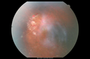
Sickle Retinpoathy SC Disease - Vitreous Hemorrhage Left Eye - 29 Year old592 views
|
|

Posterior Uveal Effusion592 views
|
|
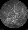
Hairy Cell Leukemia - Retinal Hemorrhage and twig Branch Vein Occlusion592 views79 year old man He has had hairy cell leukemia since 2002. He is in remission. His last blood tests were 9/2018. He just moved down here and needs a new leukemia doctor. His vision is fine.
VA OD: Dcc20/25
VA OS: Dcc20/25
IOP: TP: OD:12 OS:12
|
|

Acute Posterior Multifocal Placoid Pigment Epitheliopathy - 4 years after onset592 views4 years after presentation - vision 20/80 OD 20/16 OS and there is subretinal fibrosis
|
|

Cystoid Macular Edema 10 years after Cataract Surgery592 viewsNo overt uveitis but the patient had aching in the eyes and responded to topical anti-inflammatory drops with recurrence of the CME on discontinuation of topical therapy
|
|

Branch Retinal Artery Occlusion592 viewsAcute Inferotemporal Occlusion Right Eye - FA shows delayed circulation, OCT shows edema of superficial layers of retina
|
|

Branch Retinal Arterial Occlusion Right Eye - Atheromata - Both Eyes592 views83 year old woman with no symptoms - VA 20/40 in both eyes. Right eye has ST BRAO with silver wiring. Left eye has superotemporal atheromata
|
|

Toxoplasmosis - Recurrent - Moderate Vitritis592 views32 year old woman with 20/80 vision and moderately painful red eye in the left eye only. The left eye had some redness about 10 years ago. She is a native of Sierra Leone
|
|

Group 2a Juxtafoveal Retinal Telangiectasis (type 2)591 views58-year-old man decreased vision right eye and not better since cataract surgery one year ago. OD 20/40, OS 20/40.
|
|
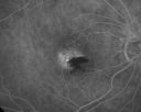
Juxtafoveal Telangiectasis Asymptomatic New Hemorrhage591 views73-year-old woman has juxtafoveal retinal telangiectasis in both eyes. The right eye has stage 5 disease. Her last Avastin treatment in the right eye was in 6 months ago. The right eye has been the worst of her two eyes for years.
VISUAL ACUITY: Vision OD is 20/200, OS is 20/50.
|
|
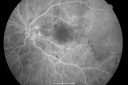
Purtchers Retinopathy with Bilateral Central Retinal Artery Occlusions 591 views50-year-old man traumatic chest compression and leg crushing injury. OD 1/200, OS 3/200
|
|

Diabetic Macular Edema Left Eye - Pre Laser591 views82-year-old woman diabetic for many years, last eye exam 5 years ago with gradual vision loss. OD 20/60, OS 20/70. IOP: OD 16, OS 17.
|
|

Diabetic Macular Edema Left Eye - Pre Laser591 views82-year-old woman diabetic for many years, last eye exam 5 years ago with gradual vision loss. OD 20/60, OS 20/70. IOP: OD 16, OS 17.
|
|

Hypertensive Retinopathy with Choroidal Ischemia Right Eye and Vision 8/200 - Blood Pressure 240/120 mmHg IgA Nephropathy591 views39-year-old man two days ago he noticed poor vision in his right eye OD: 8/200; OS: 20/20. BP is 240/120
|
|

Branch Retinal Arterial Occlusion 20/20 Vision - Symptoms for 1 Day591 views75-year-old man was seen in the office on 4/6/2010. He noticed a sudden vision change in the right eye yesterday morning. 20/20 Vision both Eyes
|
|

Idiopathic Central Serous Retinopathy - Bilateral - Acute 4 months later VA 20/40 OU591 viewsNo treatment and fluid under fovea in left eye is competely absorbed at 4 months.
|
|

Best Disease for 30 Years - 20/50 Vision both eyes - 61 Year Old Woman591 views61-year-old woman was diagnosed with Best’s disease about 30 years ago. She and her many family members were tested, both at the VA Hospital and then at USF. OD 20/50, OS 20/50
|
|

Macular Fold - 40 years post retinal detachment repair - Glaucoma 591 views79-year-old woman has had a long-standing macular fold in the right eye associated with retinal detachment repair in 1970. (40 years ago) She has a macular scar in that eye. She has optic atrophy in both eyes from glaucoma. Vision OD is hand motion, OS is 20/50
|
|

Pigmented Maculae - Mother and Daughter - Normal Visual Acuity591 views12-year-old had a dilated macular exam and the macula look abnormal. Interestingly, her mother, who is with her at this visit, was told about a year ago that her maculae are abnormal too. Her mother’s vision is fine. In the family tree, there are no known retinal dystrophies. They are both healthy.
VISUAL ACUITY: Her vision is 20/20 in both eyes.
|
|

DUSN - Diffuse Unilateral Subacute Neuroretinitis - Nematode591 views
|
|

Hyperpigmentation of Retinal Pigment Epithlium in Dry AMD (hyperplasia)591 views87-year-old woman has wet age-related macular degeneration in the left eye. She had treatment up north but there has been a gap in therapy and the vision in the left eye is poor. Vision OD is 20/25, OS is 20/400
|
|

Myopic Macular Degeneration - Staphyloma - Recent Vision Loss Right Eye - Possible CNVM591 views83-year-old woman is a high myope with myopic macular degeneration. She had a retinal detachment repair in the right eye in 1968. I saw her in June of 2003. She had posterior colobomas and areas of pigment epithelial atrophy. She has noticed over the last six weeks substantial declining vision in the right eye. Prior to that, the vision in the two eyes was about the same. She uses Xalatan in the left eye and Alphagan in both eyes.
VISUAL ACUITY: Vision OD is 20/160, OS is 20/40
|
|

Drusen Maculopathy - Macular Degeneration (Dry) - 50 year old African-American Woman591 views50-year-old woman has drusen and maculopathy. VISUAL ACUITY: OD 20/30, OS 20/25.
|
|

Choroidal Hemangioma591 views
|
|

Pattern Dystrophy - Adult Vitelliform Macular Degeneration591 views72-year-old woman 2 months post ECCE with poor vision since surgery especially left eye.
VISUAL ACUITY: OD 20/50, OS 20/200
|
|
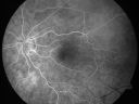
Cilioretinal artery occlusion and Ophthalmic Artery Occlusion with Relative Sparing of the Central Retinal Artery591 views80-year-old woman who says she was looking in the mirror, covered her right eye and noticed she could not see well out of the left eye. That just happened yesterday. Her last eye check was a few years ago. She also had a carotid endarterectomy on the right side in 2006 shortly after coronary bypass surgery. OD is 20/40, OS is 20/200.
|
|

Retinal Tear treated with Laser 591 views
|
|

Alphamethlyacyl COA Racemase Deficiency591 views
|
|

Angioid Streaks and Macular Degeneration - CNVM OS - Band Keratopathy 591 views79-year-old man has wet age-related macular degeneration in the left eye, for which he is having Lucentis treatment. OD 20/40, Pinhole 20/32. OS 20/160, Pinhole 20/50. He has band keratopathy sparing the visual axis in both eyes. ICG shows angioid streaks both eye.
|
|

Retinitis Pigmentosa Sine Pigmento or Unknown Dystrophy - Peripheral Superficial White Spots and Recurrent CME591 views1 month of treatment with diamox 500 mg BID- cme is a little better
|
|

Vitreomacular traction - recent vision loss - preop SD OCT591 views
|
|

Vitreo-Traction due to Diabetic Retinopathy591 viewsPatient comes in for follow up on her diabetes. VA is 20/30 in the right eye. Fundus photos shows thick traction nasally to the optic nerve forming a ring around the macula. Will follow up in 6-months.
|
|

Pre-retinal hemorrhage following Posterior Vitreous Detachment (separation - PVD)591 views68 year old woman with acute PVD OD and new preretinal hemorrhage - also there is a macular nevus
|
|

Posterior Uveal Effusion591 views
|
|
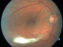
Vitreomacular traction - 20/40 vision and symptomatic with metamorphopsia591 views
|
|

Stargardt's Macular Dystrophy ABCA4 positive591 views Fundus Autofluorescence shows central atrophy with Hyper FAF pisciform triradiate lesions. Images show some progression over 3 years of macular dystrophy.
|
|

Central retinal artery and vein occlusion - Protein C Deficiency 33 year old male591 views33 year old male with vision loss for 3 hours. VA 20/12 right eye, 5/200 left eye. He has a protein C deficiency. The left eye recovered vision in about 1 month to 20/16. FA shows very slow recirculation time.
|
|
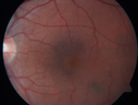
Cystoid Macular Edema 10 years after Cataract Surgery591 viewsNo overt uveitis but the patient had aching in the eyes and responded to topical anti-inflammatory drops with recurrence of the CME on discontinuation of topical therapy
|
|

Ocular Lymphoma - non-Hodgkins - B-Cell591 viewsMultifocal sub-RPE lesions in the left eye of a patient with history of Non-hodgkins B-cell Lymphoma last active 2 years ago
|
|

CRVO with Paracentral Acute Middle Maculopath - PAMM591 views56 year old woman with sudden vision loss, hemorrhages in all four quadrants. FA shows good retinal circulation. The OCT shows PAMM lesions in the affected eye. Vision did improve on second visit from 20/160 to 20/60 in about a month
|
|

Myelinated Nerve Fiber Layer - Optic Nerve591 viewsRight eye subtle and inferior - left eye obvious
|
|
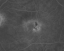
Juxtafoveal Telangiectasis - (MacTel) - Stage 4 both Eyes590 views57-year-old woman with a history of decreased vision. The patient reports that her vision has been gradually decreasing in the left more so than the right eye. 20/50 OD and 20/60
|
|
| 18117 files on 182 page(s) |
 |
|
|
|
|
52 | |
|
|
|
|


