|
|

Wet Macular Degeneration with Juxtapapillary Neovascular Membrane and Choroidal Laser Treatment572 views87-year-old woman has wet age-related macular degeneration in the left eye with an extrafoveal choroidal neovascular membrane OD is 20/25, OS is 20/25
|
|

80 Year old man with 3 day history of vision loss right eye. Vision 4/200.572 views
|
|
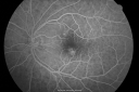
Idiopathic Central Serous Retinopathy - Bilateral - Acute 572 views49-year-old man decreasing vision in the left eye for one week. He is not taking any steroid or steroid-related products. His blood pressure as far as he knows is under control.
VISUAL ACUITY: OD 20/30, OS 20/400
|
|

Eccentric Disciform Scar - Breakthrough Vitreous Hemorrhage - InferoTemporal Subretinal Fibrosis - Ectopic Choroidal Neovascular Membranes572 views88-year-old man has noticed for the last few months the vision in the right eye is foggy, like there is a film in the eye or a haze, His vision is 20/16 in each eye.
Right eye - inferotemporal periphery there is a hemorrhage at the equator at about 7:00 o’clock, which is about 4-disc diameters across and adjacent to that there is scarring.
Left eye - large disciform scar inferotemporally
|
|

Pattern Dystrophy - Adult Vitelliform Macular Degeneration572 views72-year-old woman 2 months post ECCE with poor vision since surgery especially left eye.
VISUAL ACUITY: OD 20/50, OS 20/200
|
|

Cuticular Drusen (Basal Laminar Drusen) and Bilateral Chronic Subfoveal Fluid (Serous Retinal Detachment) unresponsive to monthly Avastin Early FA OD572 views66-year-old man with bilateral subfoveal fluid unresponsive to one year of monthly avastin therapy.
OD 20/160, Pinhole 20/80. OS 20/63, Pinhole 20/50. OCT scan shows subretinal fluid with normal choroidal thickness. Fluorescein angiogram shows cuticular/basal laminar drusen. ICG shows no foci of choriocapillaris leakage.
|
|

Retinal Detachment572 views
|
|

Choroidal Metastases - Both Eyes - Exudative Retinal Detachment572 views58-year-old man smoke a pack of cigarettes a day. He has had some ribcage pain for about the last two weeks. He also had a sinus infection for about two weeks, and he has also had hazy vision on the superior visual field in the left eye for about the last two weeks. vision is 20/25 in both eyes
|
|

Punctate Inner Choroidopathy - Recurrent CNVM 4 years after initial treatment Left Eye - Fluorescein Angiogram572 viewsJune 7, 2012. This pleasant 35-year-old woman noticed decreasing vision in the left eye recently. She returns now noticing the vision in the left eye is declining. OD 20/50, OS 20/80. OS: Vertical C/D ratio is 0.3. There is a scar just inferonasal to the fovea, which has grown some towards the fovea with a little blood just under the fovea.
|
|

Branch Retinal Artery Occlusion with Hollenhorst Plaques572 viewsBranch Retinal Artery Occlusion with Hollenhorst Plaques
|
|

Non-specific (uncharaterized / unknown) Macular Dystrophy - FAF572 views59-year-old man has macular dystrophy in both eyes. He had some vision changes in medical school in the 1980s and saw Dr. Gass for that. He had some pigment epithelial irregularities in both eyes. He had strabismus as a child and had muscle surgery. He is color blind, along with many people in his family, and as far as he knows, it is congenital.
20/25, 20/30
|
|

Primary Ocular Lymphoma - 051812 572 views
|
|

Asymptomatic Juxtapapillary Choroidal Neovascular Membrane - Reticular Macular Disease (Wet AMD) 572 views
|
|

Reticular Macular Disease (Pseudo-drusen) Both Eyes - Occult CNVM and Macular Edema Right Eye 572 viewsFluorescein Angiogram
|
|

Idiopathic Polypoidal Choroidal Vasculopathy - FAF572 views
|
|

Retinal Arterial Macroaneurysm - Untreated Hypertension (250/135) - Serous Macular Detachment572 views82 year old asian woman. VA 20/80 Headaches and vision loss for 1 month. Negative medical history but has not been to the doctor for years. (no insurance)
|
|
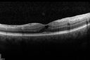
Macular Pucker 20/40 vision - minimal symptoms for 1 year Right eye572 viewsSD OCT line Scan Affected Eye
|
|

Dislocated IOL with Tube Shunt572 viewsPatient comes in with a dislocated lens in the right eye. Decreased vision in the right eye. Slit lamp photos shows lens dislocated halfway inferior. Ahmed Tube shut is visible at 11-o'clock superiorly in the right eye.
|
|
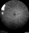
Sickle Retinopathy - Ultra Wide Field Angiogram - Heidelberg572 viewsLeakage from peripheral fronds of NV - HGB SC disease - 28 year old man with vision loss OS from acute vitreous hemorrhage
|
|

Choroidal Effusions and Serous retinal detachment with Systemic Testosterone and Cialis in 75 year old Man572 viewsFluorescein angiogram shows some staining of the disk - confocal system renders peripheral effusions black
|
|

Ocular Lymphoma - non-Hodgkins - B-Cell572 viewsMultifocal sub-RPE lesions in the left eye of a patient with history of Non-hodgkins B-cell Lymphoma last active 2 years ago
|
|
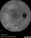
Ocular Lymphoma - non-Hodgkins - B-Cell572 viewsMultifocal sub-RPE lesions in the left eye of a patient with history of Non-hodgkins B-cell Lymphoma last active 2 years ago
|
|

Central Serous Retinopathy - Subretinal Fibrin572 views66 year old female with 20 year history of single episode of CSR. Now with vision loss for a month but improving - VA 20/63 OD; 20/32 OS
|
|

Grade 4 hypertensive retinopathy572 views55 year old man Hhe did have headaches a few weeks ago and also some nausea and vomiting. He noticed for a few weeks the vision in the left eye was blurred and that the vision in the left eye is dark.
VA OD: Dcc20/20
VA OS: Dcc20/100-1
He was admitted directly to ICU for blood pressure control (290 / 170 mmHg)
3 weeks later his VA increased to 20/20 OD, 20/32 OS
|
|

Melanosis Oculi and Choroidal Melanoma572 views88 year old female with 2.5 mm elevated choroidal melanoma and melanosis oculi. She also has geographic atrophy from AMD. The right eye is the better eye with 20/40 vision. After observation with growth, brachytherapy was done.
|
|

Wet AMD 62 year old male smoker 571 views62-year-old man has wet age-related macular degeneration in the right eye. He has been undergoing treatment with Lucentis for that. Then his left eye developed hazy vision a few weeks ago while he was overseas and now his vision is hazy in both eyes. OD 20/40, OS 20/30. FA shows a classic subfoveal choroidal neovascular membrane in the left eye, which is leaking and in the right eye, which is dry. He is going to stop smoking today. FU: VA 20/30 OU with 2 years of every 6 weeks bilateral avastin!
|
|

Wet AMD 62 year old male smoker 571 views62-year-old man has wet age-related macular degeneration in the right eye. He has been undergoing treatment with Lucentis for that. Then his left eye developed hazy vision a few weeks ago while he was overseas and now his vision is hazy in both eyes. OD 20/40, OS 20/30. FA shows a classic subfoveal choroidal neovascular membrane in the left eye, which is leaking and in the right eye, which is dry.
He is going to stop smoking today.
|
|

Cystoid Macular Edema - Xalatan - ECCE was done 20 years previously571 views71 Year old woman who noticed about a month ago the central vision in the right eye fogged up. OD 20/200 POSSIBLE XALATAN INDUCED CYSTOID MACULAR EDEMA
|
|

New BRVO Left Eye Old BRVO Right Eye - Right Eye Showing Bulls Eye Atrophy from Chronic CME and Non-Perfusion Infero-temporally571 views90-year-old man decreased vision maybe one year since cataract surgery he is no longer able to read. OD 20/200, OS 20/200
|
|

Multiple Evanescent White Dot Syndrome - Acute Idiopathic Blind Spot Enlargement571 views33-year-old woman one week ago she awoke and saw a red spots flashing out of her right eye. She notices them to the right side, which is the temporal visual field out of the right eye, then they changed to bursts of white lights without colors and then she notices straight ahead a couple spots. She has headaches in the back of her head. OD 20/20, OS 20/20
|
|

Angioid Streaks - New Extrafoveal Choroidal Neovascular Membrane Right Eye571 views61-year-old woman twelve days ago, saw a spot to the right of her central vision. OD is 20/25, OS is 20/25
Treated with one year of avastin with good result and no vision loss.
|
|

Central Retinal Artery Occlusion less than 24 hours - 69 year old man VA light perception571 views
|
|

Branch Retinal Arterial Occlusion 20/20 Vision - Symptoms for 1 Day571 views75-year-old man was seen in the office on 4/6/2010. He noticed a sudden vision change in the right eye yesterday morning. 20/20 Vision both Eyes
|
|
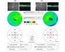
Juxtafoveal Telangiectasis - After Hemorrhage Absorbed Right Eye with Avastin Injections571 views73-year-old woman has juxtafoveal retinal telangiectasis in both eyes. The right eye has stage 5 disease. Her last Avastin treatment in the right eye was in 6 months ago. The right eye has been the worst of her two eyes for years.
VISUAL ACUITY: Vision OD is 20/200, OS is 20/50.
|
|

Basal Laminar (cuticular) Drusen both eyes 60 Year Old Woman571 views60-year-old woman with minimal vision loss. Vision OD is 20/30 OS 20/40
|
|

Myopic Macular Degeneration - Staphyloma - Recent Vision Loss Right Eye - Possible CNVM571 views83-year-old woman is a high myope with myopic macular degeneration. She had a retinal detachment repair in the right eye in 1968. I saw her in June of 2003. She had posterior colobomas and areas of pigment epithelial atrophy. She has noticed over the last six weeks substantial declining vision in the right eye. Prior to that, the vision in the two eyes was about the same. She uses Xalatan in the left eye and Alphagan in both eyes.
VISUAL ACUITY: Vision OD is 20/160, OS is 20/40
|
|

Retinal Arterial Macroaneurysm - Ruptured and self-sealed after hemorrhage571 views90-year-old woman has vision loss in the right eye for 6 weeks. OD 8/200, OS 20/50. Initial Visit
|
|

Central Retinal Vein Occlusion and Tractional Retinal Detachment for 3 Years (Patient is Diabetic)571 views67-year-old woman has severe vision loss in the left eye for 3 years OD 20/32, OS 3/200
|
|

Macular Hole - Stage 4571 views53-year-old woman was seen in the office on 5/20/2011. She had cataract surgery in June of 2009. She noticed the vision in the left eye was starting to get foggy some time around the middle of April (1 month ago).
VISUAL ACUITY: Vision OD is 20/20, OS is 20/160
|
|

Cytomegalovirus Retinitis571 viewsPatient who is HIV Positive with Cytomegalovirus Retinitis. Patient was treated with Gangcyclovir Intra-Vitreal injection.
|
|

Pre-proliferative Diabetic Retinopathy - Peripheral non-perfusion visible on FA571 views
|
|

Gonioscopy of Blood in the Angle571 viewsYoung female who has a history of uveitis, glaucoma and juvenile rheumatoid arthritis. VA is hand motion in the left eye. Pigment changes with some blood in the anterior chamber seen here on Gonioscopy.
|
|
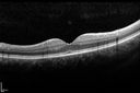
Macular Pucker 20/40 vision - minimal symptoms for 1 year Right eye571 viewsSD OCT line Scan Fellow Eye 20/16
|
|

Uveitis and Cystoid Macular Edema - Probably Sarcoidosis571 viewsVA 20/63 OD 20/50 OS - 3 months vision loss - second episode by history of uveitis
|
|

Posterior Uveal Effusion571 views
|
|

571 views
|
|

Retinal Atrophy 1 year following Streptococcus sanguinis endophthalmitis - Vision 5/200571 views79 year old woman with sleep apnea who sleeps with CPAP machine
|
|
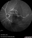
Ischemic BRVO with non-perfusion (nonperfusion) - recovered from severe vitreous hemorrhage571 views
|
|

Cystoid macular edema 4 months following cataract surgery with serous retinal detachment571 viewsCME which is chronic will often cause a serous reitnal detachment because of loss of the integrity of the external limiting membrane
|
|

Choroidal Folds Hyperope (+5.00)571 views
|
|
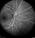
Birdshot Chorioretinitis - Fresh Presentation571 viewsUltawidefield ICG shows best the multifocal lesions. 61 year old woman with floaters for less than 6 months. HLA-A29 Positive
|
|
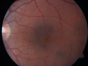
Cystoid Macular Edema 10 years after Cataract Surgery571 viewsNo overt uveitis but the patient had aching in the eyes and responded to topical anti-inflammatory drops with recurrence of the CME on discontinuation of topical therapy
|
|

Branch Retinal Artery Occlusion571 viewsAcute Inferotemporal Occlusion Right Eye - FA shows delayed circulation, OCT shows edema of superficial layers of retina
|
|

CRVO with Paracentral Acute Middle Maculopath - PAMM571 views56 year old woman with sudden vision loss, hemorrhages in all four quadrants. FA shows good retinal circulation. The OCT shows PAMM lesions in the affected eye. Vision did improve on second visit from 20/160 to 20/60 in about a month
|
|

Moderate PDR571 views43 year old man 20/32 vision in both eyes. Being started on Anti-VEGF injections which will be followed by PRP.
|
|

Macular Hole Closed with Vitrectomy - Preop570 views
|
|

Stargart's Macular Dystrophy Severe570 views32-year-old man carries a diagnosis of Stargardt’s disease with poor vision in both eyes since he was 13. He has not noticed any substantial change since he was here 4 years ago.
VISUAL ACUITY: OD 10/200, OS 8/200
|
|

Choroidal Melanoma Medium570 views33-year-old man with a history of decreased in the right eye. He had blurring in the eye about a year ago. He reports that it improved spontaneously until it was “nearly resolvedâ€. He said he had some persistent distortion, but that over the past couple of weeks he has noticed this dark spot recurring in the right eye, the left eye is unaffected. He does not have any flashes, floaters or change in his peripheral visual acuity. 20/80 OD, 20/16 OS. No improvement with pinhole. IOP: 12 OD, 11 OS.
POSTERIOR SEGMENT EXAMINATION: Shows a clear view with no PVD in either eye. C/D ratio is 0.2 OU. Large retinal vessels are unremarkable. The macula of the right eye has a brown choroidal lesion with pigment overlying the surface and orange spots along its lateral surface. The left macula is normal.
B-SCAN: B-scan ecography of the ultrasound of the right eye shows a mass about 5 mm high with a moderate to low internal reflectivity on A-scan echogram. There is serous fluid inferiorly on the echogram
|
|

Central Retinal Vein Occlusion and Cystoid Macular Edema - Acute - Vision 20/50 570 views68-year-old man six weeks of vision loss right eye from CRVO and CME, vision right eye 20/50
|
|

Central Retinal Vein Occlusion - New- and old Macular Scar from AMD570 views87-year-old woman with longstaning macular scars and new vision loss left eye for 2 months. OD: 20/200, OS 20/400.
|
|
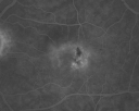
Juxtafoveal Telangiectasis - (MacTel) - Stage 4 both Eyes570 views57-year-old woman with a history of decreased vision. The patient reports that her vision has been gradually decreasing in the left more so than the right eye. 20/50 OD and 20/60
|
|

Juxtafoveal Telangiectasis - After Hemorrhage Absorbed Right Eye with Avastin Injections570 views73-year-old woman has juxtafoveal retinal telangiectasis in both eyes. The right eye has stage 5 disease. Her last Avastin treatment in the right eye was in 6 months ago. The right eye has been the worst of her two eyes for years.
VISUAL ACUITY: Vision OD is 20/200, OS is 20/50.
|
|

Hyperpigmentation of Retinal Pigment Epithlium in Dry AMD (hyperplasia)570 views87-year-old woman has wet age-related macular degeneration in the left eye. She had treatment up north but there has been a gap in therapy and the vision in the left eye is poor. Vision OD is 20/25, OS is 20/400
|
|

Hyperpigmentation of Retinal Pigment Epithlium in Dry AMD (hyperplasia)570 views87-year-old woman has wet age-related macular degeneration in the left eye. She had treatment up north but there has been a gap in therapy and the vision in the left eye is poor. Vision OD is 20/25, OS is 20/400
|
|

Nanopthalmous (Microphthalmous) Chorioretinal Folds and Choroidal Thickening Both Eyes570 views67-year-old woman with microphthalmos/nanophthalmos Her vision has gradually been getting a little bit worse, especially in the right eye. Also the anterior chamber is getting crowded and there is concern about the need for cataract surgery. She has one sister and one cousin who also have nanophthalmos.
VISUAL ACUITY: OD 20/50, OS 20/80.
|
|

Radiation Retinopathy and Macular Pucker 4 years following Brachytherapy (Plaque) for Nasal Melanoma570 views67-year-old woman has a macular pucker in the left eye. She had radiation for a melanoma in that eye 4 years aog. She had cataract surgery 2 years ago and a posterior sub-Tenon Kenalog injection in 7 months ago. Her vision in the left eye is hazy and she has trouble seeing out of it. VA OS 20/70.
|
|
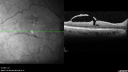
Macular Pucker - Spontaneously Partial Peel after 20 Years. Vision improved to 20/30570 views83-year-old woman decreased vision in the right eye for about a month. 20 years ago she was diagnosed with a macular pucker OD 20/50, OS 20/20.
|
|

570 views63-year-old man diabetic for twenty two years. His central vision is fine.
VISUAL ACUITY: OD 20/25, OS 20/25
|
|

Lumigan (Bimatoprost) Cystoid Macular Edema - Eye Better 1 month after Stopping Lumigan570 viewsVision back to 20/20 1 month after stopping Lumigan
|
|

Choroidal Metastases - Both Eyes - Exudative Retinal Detachment570 views58-year-old man smoke a pack of cigarettes a day. He has had some ribcage pain for about the last two weeks. He also had a sinus infection for about two weeks, and he has also had hazy vision on the superior visual field in the left eye for about the last two weeks. vision is 20/25 in both eyes
|
|

Retinitis Pigmentosa Sine Pigmento or Unknown Dystrophy - Peripheral Superficial White Spots and Recurrent CME570 views46-year-old woman three of her 5 siblings have been diagnosed with retinitis pigmentosa. Her mother and father were seen and as far as everyone can tell, neither of them has it, although there was some thought they might be carriers. They were evaluated twenty five years ago and since then as far as she knows, there has not been a lot of testing done.
She has noticed a substantial decline in vision in both eyes over the last month or two. OD 20/40, OS 20/50. IOP: Both eyes have CME
|
|

Wet AMD - New Small Hemorrhage Both Eyes - Mature CNVM visible on Early Indocyanine Green Angiogram570 views
|
|

Proliferative Diabetic Retinopathy and Vitreous Hemorrhage - FA shows leakage of dye from NVE570 views
|
|

early phase FA570 views50+ YO female presented with DM type 1;
PT presented with PDR s/p extensive PRP
FA showed CNV OD
VA stable at 20/150 for 1 year
|
|

Macular Hole Left Eye - progression from VMT - Stage II macular hole - Spontaneously Closed Macular Hole570 viewsMacular hole is closing and vision has improved to 20/40
|
|

Sickle Retinpoathy SC Disease - Vitreous Hemorrhage Left Eye - 29 Year old570 views
|
|

Choroideremia - Female Carrier - Patchy Atrophy of Choroid and RPE - Lyonization570 views
|
|

Posterior Uveal Effusion570 views
|
|

OHS CNVM post treatment with Lucentis - VA now 20/25570 views
|
|

Retinal Atrophy 1 year following Streptococcus sanguinis endophthalmitis - Vision 5/200570 views79 year old woman with sleep apnea who sleeps with CPAP machine
|
|

hydroxychloroquine (Plaquenil) Toxicity - Taiwanese 56 year old female (Asian)570 views56 year old female on 400 mg plaquenil for 22 years presents with vision loss. VA 20/50 both eyes. Plaquenil was stopped. The vision in the left eye continued to deteriorate to 3/200. 5'2" 127 lb (6.6 mg/kg body weight)
|
|

Cystoid Macular Edema 10 years after Cataract Surgery570 viewsNo overt uveitis but the patient had aching in the eyes and responded to topical anti-inflammatory drops with recurrence of the CME on discontinuation of topical therapy
|
|

Optic Nerve Pit Retinal Detachment - SD OCT of Nerve EDI shows Pit570 views9 Year old with morning glory disc who developed nerve pit RD. Raster scans of the optic nerve on EDI setting show the communication between the subretinal space and the fluid around the cerebrospinal fluid surrounding the optic nerve sheath.
|
|

Ocular Lymphoma - non-Hodgkins - B-Cell570 viewsMultifocal sub-RPE lesions in the left eye of a patient with history of Non-hodgkins B-cell Lymphoma last active 2 years ago
|
|

Branch Retinal Vein Occlusion (BRVO) no macular edema - cotton wool spot570 views85 year old man with asymptomatic BRVO - his vision stayed great without needing any treatment.
|
|

Torpedo Maculopathy570 viewsLikely a variant of CHRPE
|
|

Retinal Capillary Hemangioma - Endophytic and Exophytic570 views46 year old female with normal vision - her father died of pancreatic cancer
|
|

Cardiac Catheterization induced Cilioretinal Artery Occlusion - Paracentral Acute Middle Maculopathy570 views69 year old female with sudden vision loss on completing cardiac catheterization. The SD OCT shows a PAMM lesion and the lesion looks gray on color imaging. There is delayed filling of the cilioretinal artery.
|
|

Chronic BRAO with Hemorrhages - Sclerosed Vessel570 views95 year old female with blurred vision in both eyes in for a checkup. History of perpapillary CNVM in the left eye 2 years prior not treated. History of stroke with multiple emboli 1 year ago.
Vision 20/32 OD, 20/40 OS
|
|
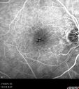
Stellate non-hereditary idiopathic foveomacular retinoschisis (SNIFR) and optic nerve drusen570 views84 year old man No visual complaints. Not diabetic, cataract surgery 8 years ago. Meds: Omeprazole, Tamsulosin (Flomax)
VA 20/32 OD, 20/20 OS
|
|

Peripheral exudative hemorrhagic chorioretinopathy - PECHR - Subfoveal fluid570 views85 year old female with
Fluctuating vision in the right eye.
VA 20/200 OD, 20/32 OS
|
|

Multiple Evanescent White Dot Syndrome - Atypical Yellow Foveal Spot Right Eye569 views12-year-old had decreased vision in the left eye from reported amblyopia since around the year 2000. His right eye was doing fine until two days ago, when he had sudden severe vision loss. He sees a blurred spot in the middle of the vision in the right eye wherever he looks.
OD 20/60 with eccentric movement, OS 20/200. Pinhole 20/70. IOP: OD 14, OS 10.
|
|

Acute Zonal Occult Outer Retinopathy with Severe Peripheral Vision Loss Left Eye 569 views 58-year-old man developed a peripheral vision problem about 25 years ago. OD 20/30, OS 20/40.
|
|

569 views
|
|
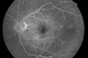
Ocular Ishemic Syndrome - Bilateral Carotid Occlusions - OD 20/60 OS 20/200 Enlarged Foveal Avascular Zone and Temporal Non-perfusion569 views75-year-old man had laryngeal cancer treated in 15 year ago with radiation. He recently was told that the carotid circulation is closed off on both sides. He does have a patent vertebral artery, which is supplying him pretty well and in addition to that his cardiac output is compromised by poor mitral valves. His consideration is to what can be done about that in the future.
VISUAL ACUITY: OD 20/60, OS 20/200
|
|

Drusen Maculopathy 85 year old Male569 views85-year-old man has age-related macular degeneration in both eyes with drusen maculopathy. OD is 20/40, OS is 20/400
|
|

Normal Fellow Eye569 views65-year-old woman has pseudoretinitis pigmentosa in the right eye only, most likely from acute zonal occult outer retinopathy. OD 20/25, OS 20/25
|
|
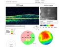
Macular Pucker 20/30 Good Vision 569 views64-year-old woman decreased vision in the left eye for about six months. You saw a wrinkle on the eye and suggested she come in here for an evaluation. She had radial keratotomy done 20 years ago.
VISUAL ACUITY: OD 20/25, OS 20/30
|
|

Nanopthalmous (Microphthalmous) Chorioretinal Folds and Choroidal Thickening Both Eyes569 views67-year-old woman with microphthalmos/nanophthalmos Her vision has gradually been getting a little bit worse, especially in the right eye. Also the anterior chamber is getting crowded and there is concern about the need for cataract surgery. She has one sister and one cousin who also have nanophthalmos.
VISUAL ACUITY: OD 20/50, OS 20/80.
|
|

Choroidal Rupture - 13 year old - 3 months following injury to eye with aluminum foil ball corneal laceration 569 views13-year-old 3 months post injury to right eye with corneal laceration and cryo to a retinal tear on July 27th and then six clock hours away laser to a retinal tear on October 20th. His vision is a little better in the eye.
VISUAL ACUITY: Vision OD is 20/120.
|
|
| 17965 files on 180 page(s) |
 |
|
|
|
|
53 | |
|
|
|
|


