|
|
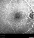
Stellate non-hereditary idiopathic foveomacular retinoschisis (SNIFR) and optic nerve drusen588 views84 year old man No visual complaints. Not diabetic, cataract surgery 8 years ago. Meds: Omeprazole, Tamsulosin (Flomax)
VA 20/32 OD, 20/20 OS
|
|

Central Retinal Vein Occlusion and Cystoid Macular Edema - Acute - Vision 20/50 587 views68-year-old man six weeks of vision loss right eye from CRVO and CME, vision right eye 20/50
|
|

Central Retinal Vein Occlusion and Cystoid Macular Edema - Acute - Vision 20/50 587 views68-year-old man six weeks of vision loss right eye from CRVO and CME, vision right eye 20/50
|
|

Idiopathic Central Serous Retinopathy - Bilateral - Acute 587 views49-year-old man decreasing vision in the left eye for one week. He is not taking any steroid or steroid-related products. His blood pressure as far as he knows is under control.
VISUAL ACUITY: OD 20/30, OS 20/400
|
|

DUSN - Diffuse Unilateral Subacute Neuroretinitis - Nematode587 views
|
|

Hyperpigmentation of Retinal Pigment Epithlium in Dry AMD (hyperplasia)587 views87-year-old woman has wet age-related macular degeneration in the left eye. She had treatment up north but there has been a gap in therapy and the vision in the left eye is poor. Vision OD is 20/25, OS is 20/400
|
|

Normal Fellow Eye587 views65-year-old woman has pseudoretinitis pigmentosa in the right eye only, most likely from acute zonal occult outer retinopathy. OD 20/25, OS 20/25
|
|
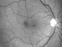
Macular Pucker - 1 year after Vitrectomy - 20/25 Vision - Postoperative Images587 views60-year-old man had vitrectomy for a macular pucker in the right eye a year ago. His vision is good.
VISUAL ACUITY: Vision OD is 20/25 Eye is now pseudophakic
|
|

Diabetic Retinopathy - Foveal Laser Scar Right Eye587 views74-year-old woman has diabetic retinopathy in both eyes. She has had focal laser in her right eye done elsewhere in the mid 90’s and unfortunately the laser was fairly close to the center and as time progressed she developed foveal atrophy around the scars. She has noticed her vision declining since she was here last a year ago. She does take Travatan in both eyes at night for glaucoma.
VISUAL ACUITY: OD 20/200, OS 20/40.
|
|
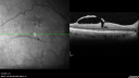
Macular Pucker - Spontaneously Partial Peel after 20 Years. Vision improved to 20/30587 views83-year-old woman decreased vision in the right eye for about a month. 20 years ago she was diagnosed with a macular pucker OD 20/50, OS 20/20.
|
|

Non-Ischemic Central Retinal Vein Occlusion in Young Man (30 years) - Many Cotton Wool Spots587 views30-year-old man with a history of a central retinal vein occlusion for which he was first examined on 2 months ago and his vision then was 4/200. He responded nicely to two Avastin treatments. OD is 20/20, OS is 20/60
|
|

Macular Hole - Stage 4587 views53-year-old woman was seen in the office on 5/20/2011. She had cataract surgery in June of 2009. She noticed the vision in the left eye was starting to get foggy some time around the middle of April (1 month ago).
VISUAL ACUITY: Vision OD is 20/20, OS is 20/160
|
|

587 views63-year-old man diabetic for twenty two years. His central vision is fine.
VISUAL ACUITY: OD 20/25, OS 20/25
|
|

Wet Age-related Macular Degeneration (AMD) Radiating Chorioretinal Folds from Contracting Macular Scar587 views93-year-old man has wet age-related macular degeneration in his left eye with a subfoveal pigment epithelial detachment.
Vision OS is 20/40. There are striking radiating chorioretinal folds emanating off of the scar, which suggests it is contracting.
|
|

Serous Retinal Detachment - Hypertensive Retinopathy - 165/120 587 views48-year-old woman noticed when she was blinking there was a white flash in the left eye starting about a month ago. She sees it temporally. OD 20/16, OS 20/20. There is a serous retinal detachment superotemporal to the optic nerve falling short of the fovea. Her blood pressure 165/120.
|
|

Medium Choroidal Melanoma Left Eye 587 views
|
|
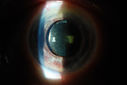
Rubeosis Iridis587 viewsPatient presents with Rubeosis Iridis in the right eye due to neovascular glaucoma. VA is 20/40 in the right eye. Will follow up in 3-months.
|
|

587 views
|
|

Idiopathic Polypoidal Choroidal Vasculopathy - FAF587 views
|
|
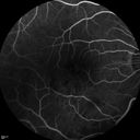
Branch Retinal Vein Occlusion with worse macular edema from hypertension - FA shows Edema Right Eye587 views
|
|

Sickle Retinopathy - Ultra Wide Field Angiogram - Heidelberg587 viewsLeakage from peripheral fronds of NV - HGB SC disease - 28 year old man with vision loss OS from acute vitreous hemorrhage
|
|

Retinal Atrophy 1 year following Streptococcus sanguinis endophthalmitis - Vision 5/200587 views79 year old woman with sleep apnea who sleeps with CPAP machine
|
|

Choroidal Folds Hyperope (+5.00)587 views
|
|
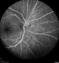
Birdshot Chorioretinitis - Fresh Presentation587 viewsUltawidefield ICG shows best the multifocal lesions. 61 year old woman with floaters for less than 6 months. HLA-A29 Positive
|
|

Coats' Disease - 14 year old - Asymptomatic587 viewsPresented with macular exudates. Temporal macroaneurysms responded to laser. Ultra-wide field FA shows nonperfusion
|
|

Hemicentral Vein Occlusion Left Eye587 viewsVision improved from 20/100 - 2 to 20/32 with 5 monthly Avastin treatments.
|
|

Ocular Lymphoma - non-Hodgkins - B-Cell587 viewsMultifocal sub-RPE lesions in the left eye of a patient with history of Non-hodgkins B-cell Lymphoma last active 2 years ago
|
|

Central Serous Retinopathy - Subretinal Fibrin587 views66 year old female with 20 year history of single episode of CSR. Now with vision loss for a month but improving - VA 20/63 OD; 20/32 OS
|
|

PAMM with Follow-up587 views
|
|
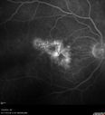
Photic Maculopathy Following Cataract Surgery587 viewsInitial images are from 1 week and subsequent images from 1 month post surgery.
|
|

Stellate Non-heredtiary Idiopathic Foveomacular Retinoschisis (SNIFR)587 views77 year old man who is healthy and 6'6" tall with 20/40 vision and no complaints. OCT shows diffuse retinoschisis.
|
|

Chronic Pseudophakic Cystoid Macular Edema in a Patient with Diabetes586 views74-year-old woman has chronic cystoid macular edema in the left eye. She has had vision loss intermittently in that eye for 2 years, the right eye, unfortunately, lost vision from a central retinal vein occlusion in 6 years ago.
|
|

Diabetic Macular Edema Left Eye - Pre Laser586 views82-year-old woman diabetic for many years, last eye exam 5 years ago with gradual vision loss. OD 20/60, OS 20/70. IOP: OD 16, OS 17.
|
|

586 views
|
|

Idiopathic Central Serous Retinopathy - Bilateral - Acute 586 views49-year-old man decreasing vision in the left eye for one week. He is not taking any steroid or steroid-related products. His blood pressure as far as he knows is under control.
VISUAL ACUITY: OD 20/30, OS 20/400
|
|

Choriodal Neovascular Membrane - Subfoveal - Pseudo-Retinitis Pigmentosa - 50 years post Retinal Detachment Repair586 views86-year-old with a history of decreased vision in the right eye. The patient reports she has lost vision in the right eye over the past two months or so. She reports the vision was always compromised in that eye since retinal detachment surgery 50 years ago with a scleral buckler. Vision dropped from 20/50 to 20/200 right eye in 2 months.
|
|

Basal Laminar (cuticular) Drusen both eyes 60 Year Old Woman586 views60-year-old woman with minimal vision loss. Vision OD is 20/30 OS 20/40
|
|

Proliferative Diabetic Retinopathy Both Eyes - Vitreous Hemorrahge Left Eye586 views67-year-old man has proliferative diabetic retinopathy in both eyes with vitreous hemorrhage in the left eye. He has had pan retinal laser in the left eye. The vitreous hemorrhage has been there for greater than two years and his vision is still hazy. He takes Lumigan in both eyes for his glaucoma.
VISUAL ACUITY: OD 20/40, OS 20/40
|
|

Choroidal Melanoma - Exudative Retinal Detachment 82 Year Old Man 586 views82-year-old man who had 3 falls 2 weeks ago. After the falls he started checking his vision and noticed there was a veil over the left eye, which he had seen for about two weeks in the superior visual field. OD 20/25, OS 20/40
(patient was medically never well enough for brachytherapy and died 6 months later from heart disease)
|
|

Radiation Retinopathy and Macular Pucker 4 years following Brachytherapy (Plaque) for Nasal Melanoma586 views67-year-old woman has a macular pucker in the left eye. She had radiation for a melanoma in that eye 4 years aog. She had cataract surgery 2 years ago and a posterior sub-Tenon Kenalog injection in 7 months ago. Her vision in the left eye is hazy and she has trouble seeing out of it. VA OS 20/70.
|
|

ECTOPIC DISCIFORM SCAR/PERIPHERAL CHOROIDAL NEOVASCULAR MEMBRANE – EXUDATE –INFLAMMATION IN REACTION TO THE PERIPHERAL VASCULAR LESION 586 views53-year-old woman has noticed some blurred vision in the right eye for a few years. In retrospect, she thinks it is possible since she was a teenager the right eye was always the weaker eye and that the left eye has always been the better eye. VISUAL ACUITY: Vision OD is 20/400
|
|

CRVO (retinal thrombophlebitis)586 views42 yr old Female c/o floaters
20/25 VA
Labs normal
|
|

Optic Nerve Hypoplasia - 43 Year Old Man - declining vision586 views43-year-old man was diagnosed with optic nerve hypoplasia at age fifteen at the National Institute of Health. His vision seems to be declining lately.
Vision OD is 20/80, OS is 20/120
|
|

FA showing NVE, NVD, non- perfusion left eye inferonasally with neovascularization at the margin of the non-perfusion586 views
|
|

Proliferative Diabetic Retinopathy and Vitreous Hemorrhage - FA shows leakage of dye from NVE586 views
|
|

Diabetic Retinopathy with CME586 viewsSecond opinion for diabetic retinopathy. VA is 20/30, right eye, 20/50, left eye. Patient has mild swelling in the macula of the right eye. No evidence of CME but does have a moderate cataract in the left eye.
|
|

AION - Acute with Follow UP RNFL scan shows edema then atrophy586 views
|
|

Hemicentral Vein Occlusion Left Eye586 viewsVision improved from 20/100 - 2 to 20/32 with 5 monthly Avastin treatments.
|
|

Hemicentral Vein Occlusion Left Eye586 viewsVision improved from 20/100 - 2 to 20/32 with 5 monthly Avastin treatments.
|
|

Asymptomatic wet AMD - Subfoveal CNM both eyes586 views82 year old woman - 20/25 OU. Images show occult subfoveal CNVM both eyes best seen on late ICG. The left eye also has probable polypoidal choroidal vasculopathy. No treatment was recommended on the first visit - follow-up at 1 month.
|
|

Retinal Capillary Hemangioma - Endophytic and Exophytic586 views46 year old female with normal vision - her father died of pancreatic cancer
|
|

Equatorial Drusen586 views61 year old female with normal visoin
|
|

Grade 4 hypertensive retinopathy586 views55 year old man Hhe did have headaches a few weeks ago and also some nausea and vomiting. He noticed for a few weeks the vision in the left eye was blurred and that the vision in the left eye is dark.
VA OD: Dcc20/20
VA OS: Dcc20/100-1
He was admitted directly to ICU for blood pressure control (290 / 170 mmHg)
3 weeks later his VA increased to 20/20 OD, 20/32 OS
|
|

Moderate PDR586 views43 year old man 20/32 vision in both eyes. Being started on Anti-VEGF injections which will be followed by PRP.
|
|

Macular Hole Closed with Vitrectomy - Preop585 views
|
|

Macular Hole Closed with Vitrectomy - Post-op 20/60 Vision585 views
|
|
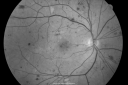
BDR and Diabetic Macular Edema with Fresh Laser Right Eye585 views60-year old man OD 20/60, OS 20/50 worsening vision both eyes
|
|

Multiple Evanescent White Dot Syndrome - Acute Idiopathic Blind Spot Enlargement585 views33-year-old woman one week ago she awoke and saw a red spots flashing out of her right eye. She notices them to the right side, which is the temporal visual field out of the right eye, then they changed to bursts of white lights without colors and then she notices straight ahead a couple spots. She has headaches in the back of her head. OD 20/20, OS 20/20
|
|

Hypertensive Choroidopathy - Pheochromocytoma - Chronic Very High Blood Pressure 49 YO Male585 views49-year-old man chronic very high blood pressure sometimes the blood pressure will go as high as 260/160, sometimes it is 200/120 and sometimes it is 160/80. He has a pheochromocytoma on the adrenal gland and he has had vision loss in the right eye starting between three and four years ago.
VISUAL ACUITY: OD 20/60, OS 20/25.
|
|

DUSN - Diffuse Unilateral Subacute Neuroretinitis - Nematode585 views
|
|

Juxtafoveal Telangiectasis - After Hemorrhage Absorbed Right Eye with Avastin Injections585 views73-year-old woman has juxtafoveal retinal telangiectasis in both eyes. The right eye has stage 5 disease. Her last Avastin treatment in the right eye was in 6 months ago. The right eye has been the worst of her two eyes for years.
VISUAL ACUITY: Vision OD is 20/200, OS is 20/50.
|
|

Nanopthalmous (Microphthalmous) Chorioretinal Folds and Choroidal Thickening Both Eyes585 views67-year-old woman with microphthalmos/nanophthalmos Her vision has gradually been getting a little bit worse, especially in the right eye. Also the anterior chamber is getting crowded and there is concern about the need for cataract surgery. She has one sister and one cousin who also have nanophthalmos.
VISUAL ACUITY: OD 20/50, OS 20/80.
|
|

Myopic Macular Degeneration - Staphyloma - Recent Vision Loss Right Eye - Possible CNVM585 views83-year-old woman is a high myope with myopic macular degeneration. She had a retinal detachment repair in the right eye in 1968. I saw her in June of 2003. She had posterior colobomas and areas of pigment epithelial atrophy. She has noticed over the last six weeks substantial declining vision in the right eye. Prior to that, the vision in the two eyes was about the same. She uses Xalatan in the left eye and Alphagan in both eyes.
VISUAL ACUITY: Vision OD is 20/160, OS is 20/40
|
|

Acute Annular Outer Retinopathy for 3 Months - 25 YO Female Fluorescein Angiogram585 views25-year-old female who, about three months ago, put on her sunglasses and noticed a smudge in the vision and at that point she noticed the right eye was hazy. She had a cold a week or two before the vision loss, but she didn’t have any fever. She has no focal neurological problems in her past history. VA 20/20 OU
|
|
585 views
|
|

Non-Ischemic Central Retinal Vein Occlusion in Young Man (30 years) - Many Cotton Wool Spots585 views30-year-old man with a history of a central retinal vein occlusion for which he was first examined on 2 months ago and his vision then was 4/200. He responded nicely to two Avastin treatments. OD is 20/20, OS is 20/60
|
|

Macular Branch Retinal Vein Occlusion and Adult Vitelliform Dystrophy585 views72-year-old woman 2 months post ECCE with poor vision since surgery especially left eye.
VISUAL ACUITY: OD 20/50, OS 20/200
|
|

Tick on Lower Lid585 viewsPt. came in for cataract surgery and a tick was found on the lower lid. Pt. had no idea the tick was attached and did not know how long it had been there. Tick was photographed and then removed by physician.
|
|
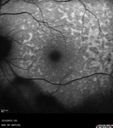
Uveitis - Retinitis - Vasculitis - possibly Syphilitis585 viewsVisit 1: 12-06-12
74-year-old man 12-3-12 visit. 2 months ago noticed vision loss, first in the right eye than the left. He is getting tired easily. He can’t walk for a long distance. He has lost five pounds within the last month. He also had a severe itchy episode, but not quite a rash a few days ago, which went away after he took a shower. FTA-ABS is positive but a spinal tap, which was negative. OD 1/200, OS 20/63.
|
|

Serous Macular Detachment Both Eyes - Wet AMD vs. CSR - 62 Year Old Man - FAF585 views
|
|

Macular Pre-Retinal Hemorrhage 585 viewsPatient complains of loss of vision in his left eye. Patient is diabetic. VA was 20/20, right eye and 20/150, left eye with no improvement pinhole. Fundus exam reveals very large pre retinal hemorrhage in the left eye. Embolism located inferior, nasally just off the optic nerve in the left eye. Patient underwent PRP for treatment of the hemorrhage.
|
|

early phase FA585 views50+ YO female presented with DM type 1;
PT presented with PDR s/p extensive PRP
FA showed CNV OD
VA stable at 20/150 for 1 year
|
|

Vitreomacular traction - normal vision on initial presentation585 views
|
|
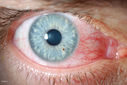
Corneal Foreign Body585 viewsPatient was cutting tile and debris flew in his eyes. Patient complained of discomfort and redness in the right eye. VA is 20/20 in the right eye. Slit lamp photography shows foreign body inferior nasal in the right eye. Removed foreign body and patched with antibiotic and will be re examined the next day.
|
|

Acute Branch Retinal Artery Occlusion Left Eye585 viewsThis 84 year old woman had an old BRVO in the right eye and saw a shadow for 2 days in the left eye. She has a superotemporal BRAO. The photos show retinal whitening, the OCT shows inner retinal edema and the FA shows poor circulation.
|
|
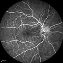
Syphillitic Optic Neuropathy and Maculopathy585 views46 year old woman with 2 weeks of vision loss OD. VA 20/200 OD; 20/20 OS. there are posterior vitreous cells on OCT in both eyes. ICG, FAF, and FA show an abnormal macula OD. VA improved to 20/30 within a month with treatment with PCN.
|
|

Syphillitic Optic Neuropathy and Maculopathy585 views46 year old woman with 2 weeks of vision loss OD. VA 20/200 OD; 20/20 OS. there are posterior vitreous cells on OCT in both eyes. ICG, FAF, and FA show an abnormal macula OD. VA improved to 20/30 within a month with treatment with PCN.
|
|

Branch Retinal Artery Occlusion585 viewsAcute Inferotemporal Occlusion Right Eye - FA shows delayed circulation, OCT shows edema of superficial layers of retina
|
|

585 views
|
|

Exogenous Fungal Endophthalmitis585 views69 year old female with endophthalmitis 2 months after ECCE. Treated with vitrectomy and 3 injections of amphotericin B. Vision improved from 5/200 - 20/50
|
|

Foveal hypoplasia - aplasia585 viewsSD OCT shows persistent plexiform layers in the center of the macula
EXTENDED HPI: He is having some trouble seeing his gadgets (like his watch). He was diagnosed with morning glory syndrome at about 3 yeras old. This last visit you saw fluid under the retina and asked him to come here. The vision is a little worse.
VA OD: Dcc20/40+2 NccJ2-2
VA OS: Dcc20/25-2 NccJ1-2
|
|

Autosomal Dominant Optic Atrophy - Father585 views52 year old man Vision has not been correctable for many years.
EXTENDED HPI: The patient has been seen for a while and his vision is not normal. Â
VA OD: sc20/100 PH20/50-1 Nsc20/400
VA OS: sc20/63 PH20/50-2 NscJ16
IOP: TP: OD:10 OS:9 Time:02:29 PM
Two siblings and his son have the same problem
|
|

Diabetic Patient with Macular Edema and Blood Pressure 200/95584 views49-year-old decreasing vision over the last year. OD is 20/80, OS 20/80. blood pressure which was 200/95.
|
|

Diabetic Retinopathy - Non-Perfusion - Possible Central Retinal Vein Occlusion584 views68-year-old woman OD 20/70, OS 20/200 CYSTOID MACULAR EDEMA, POSSIBLE EITHER OCULAR ISCHEMIC SYNDROME IN THE RIGHT EYE OR OCCULT CENTRAL RETINAL VEIN OCCLUSION IN THE RIGHT EYE,S EVERE NON-PerfUSION WITH DIABETIC RETINOPATHY IN THE LEFT EYE.
|
|

New BRVO Left Eye Old BRVO Right Eye - Right Eye Showing Bulls Eye Atrophy from Chronic CME and Non-Perfusion Infero-temporally584 views90-year-old man decreased vision maybe one year since cataract surgery he is no longer able to read. OD 20/200, OS 20/200
|
|
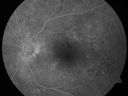
Clinically Significant Diabetic Macular Edema both Eyes with Exudates584 views56-year-old woman with diabetes for twelve years. She has noticed blurring of the vision in the right eye for about six months. OD is 20/70, OS is 20/25.
|
|

Hypertensive Choroidopathy - Pheochromocytoma - Chronic Very High Blood Pressure 49 YO Male584 views49-year-old man chronic very high blood pressure sometimes the blood pressure will go as high as 260/160, sometimes it is 200/120 and sometimes it is 160/80. He has a pheochromocytoma on the adrenal gland and he has had vision loss in the right eye starting between three and four years ago.
VISUAL ACUITY: OD 20/60, OS 20/25.
|
|

Drusen Maculopathy 85 year old Male584 views85-year-old man has age-related macular degeneration in both eyes with drusen maculopathy. OD is 20/40, OS is 20/400
|
|
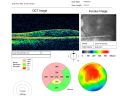
Macular Pucker 20/30 Good Vision 584 views64-year-old woman decreased vision in the left eye for about six months. You saw a wrinkle on the eye and suggested she come in here for an evaluation. She had radial keratotomy done 20 years ago.
VISUAL ACUITY: OD 20/25, OS 20/30
|
|

Acute Annular Outer Retinopathy for 3 Months - 25 YO Female Fellow Normal Eye584 views25-year-old female who, about three months ago, put on her sunglasses and noticed a smudge in the vision and at that point she noticed the right eye was hazy. She had a cold a week or two before the vision loss, but she didn’t have any fever. She has no focal neurological problems in her past history. VA 20/20 OU
|
|

Video - Takes a LONG TIME TO LOAD - X-linked Juvenille Retinoschisis - Peripheral Retinal Vascular Anomalies - Vitreous Hemorrhage - 8 Year Old Boy584 views8-year-old child OD 20/40, OS 20/50
OD: Vertical C/D ratio is 0.2. There are foveal cysts. There is also a retinal elevation inferiorly and there are patchy peripheral retinal hemorrhages.
OS: Vertical C/D ratio is 0.2. There are foveal cysts. There is peripheral retinal issues with some hemorrhage in some areas of peripheral retinoschisis.
|
|

Sickle Cell (FA)584 views
|
|

Choroidal Metastases - Both Eyes - Exudative Retinal Detachment584 views58-year-old man smoke a pack of cigarettes a day. He has had some ribcage pain for about the last two weeks. He also had a sinus infection for about two weeks, and he has also had hazy vision on the superior visual field in the left eye for about the last two weeks. vision is 20/25 in both eyes
|
|
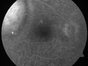
Choroidal Metastases - Both Eyes - Exudative Retinal Detachment584 views58-year-old man smoke a pack of cigarettes a day. He has had some ribcage pain for about the last two weeks. He also had a sinus infection for about two weeks, and he has also had hazy vision on the superior visual field in the left eye for about the last two weeks. vision is 20/25 in both eyes
|
|
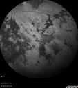
Retinitis Pigmentosa - Autosomal Dominant - Vision Loss at Young Age - Infrared584 views 54-year-old woman has autosomal dominant retinitis pigmentosa. Her mother and grandmother both had it and she has one son who has it and one daughter who doesn’t. She also has a sister with it. She lost vision since she was younger. She had cataract surgery done in January and February of this year. She has been seeing unusual flashing lights throughout the periphery and central vision for about the last year or two.
VISUAL ACUITY: OD 20/160, OS 20/160
|
|

Reticular Macular Disease (Pseudo-drusen) Both Eyes - Occult CNVM and Macular Edema Right Eye 584 viewsFundus Autofluorescence
|
|

Acute non arteritic anterior ischemic optic neuropathy left eye - SD Optical Coherence Tomography Line Scan584 views66 y.o. woman spot in central vision in left eye started about 1 week ago since then it is not changing. VA OD: sc20/25 OS: sc20/200
|
|

Branch Retinal Vein Occlusion with worse macular edema from hypertension - Photo from 2008 with initial occlusion - Vision 20/40584 views
|
|

Dislocated IOL with Tube Shunt584 viewsPatient comes in with a dislocated lens in the right eye. Decreased vision in the right eye. Slit lamp photos shows lens dislocated halfway inferior. Ahmed Tube shut is visible at 11-o'clock superiorly in the right eye.
|
|

Posterior Uveal Effusion584 views
|
|
| 18117 files on 182 page(s) |
 |
|
|
|
|
54 | |
|
|
|
|


