|
|
Photic Maculopathy Following Cataract Surgery583 x angesehenInitial images are from 1 week and subsequent images from 1 month post surgery.
|
|

Foveal hypoplasia - aplasia583 x angesehenSD OCT shows persistent plexiform layers in the center of the macula
EXTENDED HPI: He is having some trouble seeing his gadgets (like his watch). He was diagnosed with morning glory syndrome at about 3 yeras old. This last visit you saw fluid under the retina and asked him to come here. The vision is a little worse.
VA OD: Dcc20/40+2 NccJ2-2
VA OS: Dcc20/25-2 NccJ1-2
|
|
Birdshot Chorioretintis and PAMM583 x angesehenPatient presented with paracentral acute middle maculopathy as the presenting symptom of birdshot. This case was published. RETINAL CASES & BRIEF REPORTS 10:151–153, 2016.
|
|

Melanosis Oculi and Choroidal Melanoma583 x angesehen88 year old female with 2.5 mm elevated choroidal melanoma and melanosis oculi. She also has geographic atrophy from AMD. The right eye is the better eye with 20/40 vision. After observation with growth, brachytherapy was done.
|
|

Type 1 Retinal Arteriovenous Malformation583 x angesehen42 year old female VA 20/32 OD, 20/20 OS MRI was negative.
|
|

Central Retinal Vein Occlusion and Cystoid Macular Edema - Acute - Vision 20/50 582 x angesehen68-year-old man six weeks of vision loss right eye from CRVO and CME, vision right eye 20/50
|
|

582 x angesehen49-year-old woman has central retinal vein occlusion right eye with fluctuating vision for a few month now with OD 20/100. IOP: OD 20
|
|

New BRVO Left Eye Old BRVO Right Eye - Right Eye Showing Bulls Eye Atrophy from Chronic CME and Non-Perfusion Infero-temporally582 x angesehen90-year-old man decreased vision maybe one year since cataract surgery he is no longer able to read. OD 20/200, OS 20/200
|
|

Acute Zonal Occult Outer Retinopathy with Severe Peripheral Vision Loss Left Eye 582 x angesehen 58-year-old man developed a peripheral vision problem about 25 years ago. OD 20/30, OS 20/40.
|
|

Hypertensive Choroidopathy - Pheochromocytoma - Chronic Very High Blood Pressure 49 YO Male582 x angesehen49-year-old man chronic very high blood pressure sometimes the blood pressure will go as high as 260/160, sometimes it is 200/120 and sometimes it is 160/80. He has a pheochromocytoma on the adrenal gland and he has had vision loss in the right eye starting between three and four years ago.
VISUAL ACUITY: OD 20/60, OS 20/25.
|
|

Central Retinal Vein Occlusion Recurrent Edema 6 weeks after Lucentis - Nonperfusion Temporally582 x angesehen66-year-old woman has a central retinal vein occlusion in the right eye with macular edema. She had intravitreal Lucentis treatment six weeks ago and she has noticed over the last week or two her vision is declining.
VISUAL ACUITY: OD 20/70, OS 20/30.
|
|

Choroidal Rupture - 13 year old - 3 months following injury to eye with aluminum foil ball corneal laceration 582 x angesehen13-year-old 3 months post injury to right eye with corneal laceration and cryo to a retinal tear on July 27th and then six clock hours away laser to a retinal tear on October 20th. His vision is a little better in the eye.
VISUAL ACUITY: Vision OD is 20/120.
|
|

Diabetic Macular Edema582 x angesehen63 yr old Male
VA 20/20 OD 20/25 OS
No complaints
|
|

Chronic Pseudophakic Cystoid Macular Edema - Haptic Migrated into Anterior Chamber Through Peripheral Iridotomy582 x angesehen57-year-old man with congenital nystagmus. He had cataract surgery done 30 years ago and has decreased vision left eye for 6 months. OD is 20/50, OS is 20/100
|
|

582 x angesehen
|
|

582 x angesehen63-year-old man diabetic for twenty two years. His central vision is fine.
VISUAL ACUITY: OD 20/25, OS 20/25
|
|
Choroidal Metastases - Both Eyes - Exudative Retinal Detachment582 x angesehen58-year-old man smoke a pack of cigarettes a day. He has had some ribcage pain for about the last two weeks. He also had a sinus infection for about two weeks, and he has also had hazy vision on the superior visual field in the left eye for about the last two weeks. vision is 20/25 in both eyes
|
|

Retinitis Pigmentosa - Autosomal Dominant - Vision Loss at Young Age - Infrared582 x angesehen 54-year-old woman has autosomal dominant retinitis pigmentosa. Her mother and grandmother both had it and she has one son who has it and one daughter who doesn’t. She also has a sister with it. She lost vision since she was younger. She had cataract surgery done in January and February of this year. She has been seeing unusual flashing lights throughout the periphery and central vision for about the last year or two.
VISUAL ACUITY: OD 20/160, OS 20/160
|
|

FA showing NVE, NVD, non- perfusion left eye inferonasally with neovascularization at the margin of the non-perfusion582 x angesehen
|
|

Cytomegalovirus Retinitis582 x angesehenPatient who is HIV Positive with Cytomegalovirus Retinitis. Patient was treated with Gangcyclovir Intra-Vitreal injection.
|
|

Gonioscopy of Blood in the Angle582 x angesehenYoung female who has a history of uveitis, glaucoma and juvenile rheumatoid arthritis. VA is hand motion in the left eye. Pigment changes with some blood in the anterior chamber seen here on Gonioscopy.
|
|

Papilledema - Sagittal Cerebral Vein Thrombosis - Lung Transplant Cystic Fibrosis 36 Year Old Man582 x angesehenColor Photo
|
|

Branch Retinal Vein Occlusion with worse macular edema from hypertension - Photo from 2008 with initial occlusion - Vision 20/40582 x angesehen
|
|

Macular Telangiectasia - Stage 3 right eye 20/40 vision582 x angesehenConfocal InfraRed Reflectance
|
|

Branch Retinal Vein Occlusion with NVE (Neovascularization Elsewhere)582 x angesehen
|
|

Cystoid macular edema 4 months following cataract surgery with serous retinal detachment582 x angesehenCME which is chronic will often cause a serous reitnal detachment because of loss of the integrity of the external limiting membrane
|
|
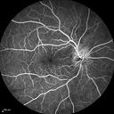
Syphillitic Optic Neuropathy and Maculopathy582 x angesehen46 year old woman with 2 weeks of vision loss OD. VA 20/200 OD; 20/20 OS. there are posterior vitreous cells on OCT in both eyes. ICG, FAF, and FA show an abnormal macula OD. VA improved to 20/30 within a month with treatment with PCN.
|
|

Syphillitic Optic Neuropathy and Maculopathy582 x angesehen46 year old woman with 2 weeks of vision loss OD. VA 20/200 OD; 20/20 OS. there are posterior vitreous cells on OCT in both eyes. ICG, FAF, and FA show an abnormal macula OD. VA improved to 20/30 within a month with treatment with PCN.
|
|

Optic Nerve Pit Retinal Detachment - SD OCT of Nerve EDI shows Pit582 x angesehen9 Year old with morning glory disc who developed nerve pit RD. Raster scans of the optic nerve on EDI setting show the communication between the subretinal space and the fluid around the cerebrospinal fluid surrounding the optic nerve sheath.
|
|

Optic Nerve Pit Retinal Detachment - SD OCT of Nerve EDI shows Pit582 x angesehen9 Year old with morning glory disc who developed nerve pit RD. Raster scans of the optic nerve on EDI setting show the communication between the subretinal space and the fluid around the cerebrospinal fluid surrounding the optic nerve sheath.
|
|
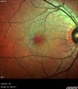
Macular Telangiectasis with Symptomatic Blind Spots582 x angesehen55 year old man with non-neovascular MacTel. VA 20/20 right eye 20/50 left eye. The color images show gray areas temporally. The FAF shows decreased macular pigment. The OCT shows outer retinal atrophy.
|
|

Branch Retinal Vein Occlusion (BRVO) no macular edema - cotton wool spot582 x angesehen85 year old man with asymptomatic BRVO - his vision stayed great without needing any treatment.
|
|

Concentric Geographic Atrophy582 x angesehen76 year old man Gradual vision loss
20/32 OD; 20/40 OS
No medicines, Non-smoker
Working and Driving
|
|

Diabetic Retinopathy - Non-Perfusion - Possible Central Retinal Vein Occlusion581 x angesehen68-year-old woman OD 20/70, OS 20/200 CYSTOID MACULAR EDEMA, POSSIBLE EITHER OCULAR ISCHEMIC SYNDROME IN THE RIGHT EYE OR OCCULT CENTRAL RETINAL VEIN OCCLUSION IN THE RIGHT EYE,S EVERE NON-PerfUSION WITH DIABETIC RETINOPATHY IN THE LEFT EYE.
|
|

80 Year old man with 3 day history of vision loss right eye. Vision 4/200.581 x angesehen
|
|

Birdshot Chorioretinitis - Retinal Vasculitis - HLA A29 Positive 581 x angesehen64-year-old woman three or four year’s fluctuations in her vision. She mostly notices them in bright light her vision glares out and she has difficulty seeing. PMHx: malaria in 1948 that was treated. She was told at one point she could possibly have had Familial Mediterranean fever.
She does intermittently get rashes around her arms. She has arthritis in her knees and ankles.
VISUAL ACUITY: OD 20/40, OS 20/50 - HLA - A29 Positive
|
|

Birdshot Chorioretinitis - Retinal Vasculitis - HLA A29 Positive 581 x angesehen64-year-old woman three or four year’s fluctuations in her vision. She mostly notices them in bright light her vision glares out and she has difficulty seeing. PMHx: malaria in 1948 that was treated. She was told at one point she could possibly have had Familial Mediterranean fever.
She does intermittently get rashes around her arms. She has arthritis in her knees and ankles.
VISUAL ACUITY: OD 20/40, OS 20/50 - HLA - A29 Positive
|
|

Proliferative Sickle Retinopathy - Hemoglobin SC581 x angesehen53-year-old woman with a new spider-like floater in the right eye for about six days. She sees one big floater and another little one. She does have hemoglobin SC hemoglobinopathy. VISUAL ACUITY: Vision OD is 20/20; OS is 20/50, PH is 20/20.
|
|

Macular Pucker - 3 Weeks Vision Loss - 20/50 Vision - Preoperative Images581 x angesehenPREOP:
59-year-old man new onset floaters about a year ago. Subsequent to that his vision was not quite right in the right eye and then he noticed, starting about three weeks ago, substantial change in the vision. VISUAL ACUITY: Vision OD is 20/50, OS is 20/16
|
|
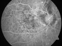
Angioid Streaks - Pseudoxanthoma Elasticum - Supressed CNVM581 x angesehen54-year-old woman had angioid streaks and macular degeneration in both eyes. She has had intravitreal Avastin in the right eye about every three months and her vision in that eye is good.
VISUAL ACUITY: Vision OD is 20/30, OS is 2/200
|
|

Basal Laminar Drusen - Cuticular Drusen - Equatorial Drusen - 70 Year Old Woman - Fundus Photo Left Eye581 x angesehen70 year old woman has had macular drusen since her thirties and she said her sister has a similar problem. Recently the vision in the left eye has substantially declined. OD 20/16, OS 10/200.
|
|

Macular Branch Retinal Vein Occlusion and Adult Vitelliform Dystrophy581 x angesehen72-year-old woman 2 months post ECCE with poor vision since surgery especially left eye.
VISUAL ACUITY: OD 20/50, OS 20/200
|
|

Video - Takes a LONG TIME TO LOAD - X-linked Juvenille Retinoschisis - Peripheral Retinal Vascular Anomalies - Vitreous Hemorrhage - 8 Year Old Boy581 x angesehen8-year-old child OD 20/40, OS 20/50
OD: Vertical C/D ratio is 0.2. There are foveal cysts. There is also a retinal elevation inferiorly and there are patchy peripheral retinal hemorrhages.
OS: Vertical C/D ratio is 0.2. There are foveal cysts. There is peripheral retinal issues with some hemorrhage in some areas of peripheral retinoschisis.
|
|

Lumigan (Bimatoprost) Cystoid Macular Edema - Eye Better 1 month after Stopping Lumigan581 x angesehenVision back to 20/20 1 month after stopping Lumigan
|
|

Vasculitis - Retinitis - Uveitis - Vision NLP right eye , 20/50 left eye581 x angesehen
|
|
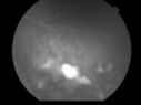
Uveitis and Retinal Whitening - 04-27-12 - Prior to PPV OD - Possible Primary Ocular Lymphoma 581 x angesehen
|
|
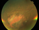
Primary Ocular Lymphoma - 051812 581 x angesehen
|
|

581 x angesehen
|
|

Pseudoxanthoma Elasticum with Angioid Streaks581 x angesehenYoung female patient who comes in for routine eye exam, VA 20/20 in both eyes. Fundus photos shows a Peu'd orange appearance in the periphery of the retina and angioid streaks around the optic nerve.
|
|

Uveitis and Cystoid Macular Edema - Probably Sarcoidosis581 x angesehenVA 20/63 OD 20/50 OS - 3 months vision loss - second episode by history of uveitis
|
|

Retinitis Pigmentosa581 x angesehenPatient comes in with cloudy vision. VA is 20/200 in both eyes. Patient has had retinal pigmentosa since 1986. Patient to consider cataract surgery to help her visual symptoms...
|
|
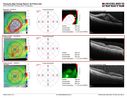
Self Sealed Retinal Arteriol Macroaneurysm - MAP shows decreased swelling581 x angesehenSD OCT map
|
|
Posterior Uveal EffusionPosterior Uveal Effusion581 x angesehen
|
|

Presumed Ocular Histoplasmosis with new CNVM in right and better eye - VA 20/80 OD; 20/800 OS581 x angesehen
|
|
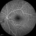
Choroidal Folds Hyperope (+5.00)581 x angesehen
|
|

Syphilitic Chorioretinitis581 x angesehenSevere outer retinal atrophy and RPE non-geographic atrophy from syphilis. Vision improved from 20/63 to 20/32 with therapy. Images look a lot like DUSN. Patient had several surgeries (vitrectomy for floaters, vitrectomy for macular pucker, Avastin injections and macular laser for edema) prior by other retina specialists prior to being correctly diagnosed and treated.
|
|

Hemicentral Vein Occlusion Left Eye581 x angesehenVision improved from 20/100 - 2 to 20/32 with 5 monthly Avastin treatments.
|
|

CRVO with Paracentral Acute Middle Maculopath - PAMM581 x angesehen56 year old woman with sudden vision loss, hemorrhages in all four quadrants. FA shows good retinal circulation. The OCT shows PAMM lesions in the affected eye. Vision did improve on second visit from 20/160 to 20/60 in about a month
|
|

Multifocal Bilateral Central Serous Retinopathy in African American Male581 x angesehen67 year old african american male with : Blurred Vision OD. Duration of Problem: 4-6 weeks. VA 20/32 OD 20/20 OS
|
|

Macular Laser Scars581 x angesehen86 year old man who had Laser done from 2010 - 2018 for diabetic retinopathy. The right eye lost vision immediately after laser was done a few years ago. VA 1/200 OD; 20/25 OS
|
|

Macular Laser Scars581 x angesehen86 year old man who had Laser done from 2010 - 2018 for diabetic retinopathy. The right eye lost vision immediately after laser was done a few years ago. VA 1/200 OD; 20/25 OS
|
|

Multiple Evanescent White Dot Syndrome - Atypical Yellow Foveal Spot Right Eye580 x angesehen12-year-old had decreased vision in the left eye from reported amblyopia since around the year 2000. His right eye was doing fine until two days ago, when he had sudden severe vision loss. He sees a blurred spot in the middle of the vision in the right eye wherever he looks.
OD 20/60 with eccentric movement, OS 20/200. Pinhole 20/70. IOP: OD 14, OS 10.
|
|

580 x angesehen66-year-old woman had a gunshot wound to the head on August 12, 2010, with the entry wound just on the right side, just about below her temple, toward the lower part of the temple and the exit wound on the left side just above her cheek, a little behind the eye. Since then she recovered and did not need any surgery. When that all cleared she realized her vision was poor in both eyes. She has seen a neuro-ophthalmologist who thought she had traumatic optic neuropathy. OD light perception, OS 20/400.
|
|
Perforating Steel Tip Dart Injury - 1 year later - no vitrectomy - 20/40 Vision 580 x angesehen16-year-old had a dart injury to the left eye one year ago. His vision is improving remarkably.
VISUAL ACUITY: OS: 20/40
|
|
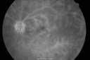
Birdshot Chorioretinitis - Retinal Vasculitis - HLA A29 Positive 580 x angesehen64-year-old woman three or four year’s fluctuations in her vision. She mostly notices them in bright light her vision glares out and she has difficulty seeing. PMHx: malaria in 1948 that was treated. She was told at one point she could possibly have had Familial Mediterranean fever.
She does intermittently get rashes around her arms. She has arthritis in her knees and ankles.
VISUAL ACUITY: OD 20/40, OS 20/50 - HLA - A29 Positive
|
|

Acute Annular Outer Retinopathy for 3 Months - 25 YO Female Fellow Normal Eye580 x angesehen25-year-old female who, about three months ago, put on her sunglasses and noticed a smudge in the vision and at that point she noticed the right eye was hazy. She had a cold a week or two before the vision loss, but she didn’t have any fever. She has no focal neurological problems in her past history. VA 20/20 OU
|
|

Malignany Hypertension - BP 250/130 - Vision 1/200 in each eye - Diabetic580 x angesehen68-year-old woman poor vision in both eyes for the last few years. She is diabetic since 1989. Her blood pressure has been very high. She was in the hospital Friday. Her blood pressure during that visit was 250/130 and her blood pressure in your office she reports was somewhere around the same. I just checked her blood pressure at the beginning of my visit and it was 200/120 in her right arm sitting. Her vision is poor enough that she can barely see to walk around. She has also been feeling nauseous
|
|
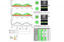
Congenital Achromatopsia 580 x angesehen26-year-old man was diagnosed with congenital achromatopsia by ERG as a child. He has family members who also have this disorder. OD 20/200, OS 20/200.
|
|
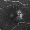
Group 2a Juxtafoveal Telangiectasis - MacTel - Stage 5 left eye - Macular Hole Right eye580 x angesehen76-year-old woman has juxtafoveal retinal telangiectasis. She had Stage five neovascularization in the left eye I treated with injections between September of 2007 and October of 2008 with Avastin (3 years ago). OCT scan today shows macular hole right eye. VISUAL ACUITY: OD 20/400, OS 20/40.
|
|

Sickle Cell (FA)580 x angesehen
|
|
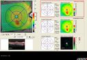
Diabetic Macular Edema - Mild580 x angesehen70 year old man with mild edema did well without treatment
|
|

Choroidal Metastases - Both Eyes - Exudative Retinal Detachment580 x angesehen58-year-old man smoke a pack of cigarettes a day. He has had some ribcage pain for about the last two weeks. He also had a sinus infection for about two weeks, and he has also had hazy vision on the superior visual field in the left eye for about the last two weeks. vision is 20/25 in both eyes
|
|

Diabetic Papillitis580 x angesehenPatient is about to undergo surgery for CNS Aneurysm. Patient woke up with little spots which never cleared up. Patients VA was 20/30-OD and 20/40-OS. Both eyes appeared to have disc edema with hemorrhages in the right eye. Ordered a CT of the brain to make sure the aneurysm didn't ruptured.
|
|

Central Serous Retinopathy 25 year old professional baseball player - Atypical - Choroidal Hypo-perfusion?580 x angesehen
|
|
Primary Ocular Lymphoma - 051812 580 x angesehen
|
|
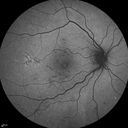
FAF580 x angesehen
|
|

Medium Choroidal Melanoma Left Eye 580 x angesehen
|
|

Acute non arteritic anterior ischemic optic neuropathy left eye - SD Optical Coherence Tomography Line Scan580 x angesehen66 y.o. woman spot in central vision in left eye started about 1 week ago since then it is not changing. VA OD: sc20/25 OS: sc20/200
|
|

Papilledema unspecified 580 x angesehen
|
|

Vitreomacular traction - recent vision loss Right Eye - initially worse after Jetrea580 x angesehen
|
|

Branch Retinal Vein Occlusion with NVE (Neovascularization Elsewhere)580 x angesehenSD-OCT of NVE - shows it to be pre-retinal
|
|

Unilateral Acute Idiopathic Maculopathy (UAIM)580 x angesehen
|
|

Unilateral Acute Idiopathic Maculopathy (UAIM)580 x angesehen41 year old with serous retinal detachment OS and sudden vision loss. He had a viral prodrome. Coxscackie IgG was high but no acute infection was documented. Vision improved in about 2 months without therapy.
|
|
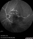
Ischemic BRVO with non-perfusion (nonperfusion) - recovered from severe vitreous hemorrhage580 x angesehen
|
|

Hypertensive Retinopathy - BP 240 / 130 mmHg - Grade 4580 x angesehen71 year old woman who claims to be asymptomatic with decreased vision, macular edema, cotton wool spots, subretinal fluid and optic nerve edema
|
|

Coats' Disease - 14 year old - Asymptomatic580 x angesehenPresented with macular exudates. Temporal macroaneurysms responded to laser. Ultra-wide field FA shows nonperfusion
|
|

Macular Atrophy - Phototoxicity - Macular Hole Surgery580 x angesehen25 years ago patient suffered immediate severe vision loss following macular hole surgery. You can almost see light pipe tracks in the atrophy in the macula. Vision is 5/200
|
|

PDR NVD left eye and NVE right eye580 x angesehen41 year old diabetic woman with PDR in both eyes. High risk in the left eye. She had PRP, Avastin, and vitrectomy x 2 in the left eye with final vision of 20/25. The right eye had eventually PRP laser
|
|

Choroidal Osteoma 13 Year Old580 x angesehen13 year old with 6 months of vision loss right eye and vision of 20/40
|
|

Sarcoidosis Multifocal Choroiditis580 x angesehen66 year old man with dense cataracts and recurrent uveitis. Images show multifocal choroidal granulomas from sarcoidosis more in the right eye than the left eye.
|
|

579 x angesehen33-year-old man with a history of decreased in the right eye. He had blurring in the eye about a year ago. He reports that it improved spontaneously until it was “nearly resolvedâ€. He said he had some persistent distortion, but that over the past couple of weeks he has noticed this dark spot recurring in the right eye, the left eye is unaffected. He does not have any flashes, floaters or change in his peripheral visual acuity. 20/80 OD, 20/16 OS. No improvement with pinhole. IOP: 12 OD, 11 OS.
POSTERIOR SEGMENT EXAMINATION: Shows a clear view with no PVD in either eye. C/D ratio is 0.2 OU. Large retinal vessels are unremarkable. The macula of the right eye has a brown choroidal lesion with pigment overlying the surface and orange spots along its lateral surface. The left macula is normal.
B-SCAN: B-scan ecography of the ultrasound of the right eye shows a mass about 5 mm high with a moderate to low internal reflectivity on A-scan echogram. There is serous fluid inferiorly on the echogram
|
|
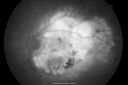
Disciform Macular Scar Both Eyes from Wet Macular Degeneration579 x angesehen86-year-old woman has disciform scars OD: 20/400; OS: 20/100
|
|

Retinal Arterial Atheromata579 x angesehen68-year-old woman with records going back 7 years, which clearly document retinal arterial plaques in both eyes.
|
|

579 x angesehen
|
|

Acute Zonal Occult Outer Retinopathy with Severe Peripheral Vision Loss Left Eye 579 x angesehen 58-year-old man developed a peripheral vision problem about 25 years ago. OD 20/30, OS 20/40.
|
|

Wet Macular Degeneration with Juxtapapillary Neovascular Membrane and Choroidal Laser Treatment579 x angesehen87-year-old woman has wet age-related macular degeneration in the left eye with an extrafoveal choroidal neovascular membrane OD is 20/25, OS is 20/25
|
|

Hypertensive Choroidopathy - Pheochromocytoma - Chronic Very High Blood Pressure 49 YO Male579 x angesehen49-year-old man chronic very high blood pressure sometimes the blood pressure will go as high as 260/160, sometimes it is 200/120 and sometimes it is 160/80. He has a pheochromocytoma on the adrenal gland and he has had vision loss in the right eye starting between three and four years ago.
VISUAL ACUITY: OD 20/60, OS 20/25.
|
|

DUSN - Diffuse Unilateral Subacute Neuroretinitis - Nematode579 x angesehen
|
|

Macular Pucker 20/40 vision and recent cataract surgery579 x angesehen 73-year-old man distortion and vision loss since ECCE 6 months ago OS. Patient taking anti-inflammatory drops. VISUAL ACUITY: OD 20/30, OS 20/40.
|
|

Acute Annular Outer Retinopathy for 3 Months - 25 YO Female FA579 x angesehen25-year-old female who, about three months ago, put on her sunglasses and noticed a smudge in the vision and at that point she noticed the right eye was hazy. She had a cold a week or two before the vision loss, but she didn’t have any fever. She has no focal neurological problems in her past history. VA 20/20 OU
|
|
| 18117 Dateien auf 182 Seite(n) |
 |
|
|
|
|
55 | |
|
|
|
|


