|
|

Asymptomatic Juxtapapillary Choroidal Neovascular Membrane - Reticular Macular Disease (Wet AMD) 580 views
|
|

Pigmented Peripheral Retinal Degeneration580 views42-year old male comes in for routine eye exam and to follow up on peripheral retinal degeneration in both eyes. VA is 20/20, right eye and 20/25, left eye. Patient is asymptomatic with no visual complaints.
|
|
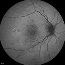
FAF580 views
|
|

Medium Choroidal Melanoma Left Eye 580 views
|
|

Chronic Central Serous Retinopathy - Indocyanine Green Angiogram 580 views
|
|

Papilledema unspecified 580 views
|
|

Cartwheel CNVM - Classic - wet AMD - evidence of possible old CSR580 views75 y.o Decreased Vision right eye for one year. VA OD: sc20/125+2 OS 20/25. Autofluorescence and SD OCT show evidence of old serous retinal detachment superior to the fovea suggesting a history of CSR. The macular lesion on FA shows a classic cartwheel appearance
|
|

Unilateral Acute Idiopathic Maculopathy (UAIM)580 views41 year old with serous retinal detachment OS and sudden vision loss. He had a viral prodrome. Coxscackie IgG was high but no acute infection was documented. Vision improved in about 2 months without therapy.
|
|

Giant Retinal Tear Retinal Detachment580 views13 year old african american girl high myope. These intra-operative photos show parts of the procedure for this giant tear detachment. The video is on youtube. You can see the center of the macula with no overlying retina.
|
|

Hypertensive Retinopathy - BP 240 / 130 mmHg - Grade 4580 views71 year old woman who claims to be asymptomatic with decreased vision, macular edema, cotton wool spots, subretinal fluid and optic nerve edema
|
|

Macular Atrophy - Phototoxicity - Macular Hole Surgery580 views25 years ago patient suffered immediate severe vision loss following macular hole surgery. You can almost see light pipe tracks in the atrophy in the macula. Vision is 5/200
|
|

PDR NVD left eye and NVE right eye580 views41 year old diabetic woman with PDR in both eyes. High risk in the left eye. She had PRP, Avastin, and vitrectomy x 2 in the left eye with final vision of 20/25. The right eye had eventually PRP laser
|
|

Recurrent MEWDS Left Eye - Teenage Girl580 viewsPatient presented in 2014 (first images) 2 months following vision loss left eye. Vision went from 20/60 - 20/20. OCT showed outer retinal irregularities which normalized. Then 3 years later in 2017 she had full blown MEWDS. She presented acutely and all the images are typical. Both episodes where in the same time of year, about 3 months after her flu shot for sports.
|
|

Choroidal Osteoma 13 Year Old580 views13 year old with 6 months of vision loss right eye and vision of 20/40
|
|

Autoimmune Retinopathy580 views91 year old female with severe vision loss over the past 3 years. She has shimmery scotomas. The main findings are outer retinal atrophy evident on OCT and FAF. VA 20/80 OD and 20/125 OS
|
|

579 views33-year-old man with a history of decreased in the right eye. He had blurring in the eye about a year ago. He reports that it improved spontaneously until it was “nearly resolvedâ€. He said he had some persistent distortion, but that over the past couple of weeks he has noticed this dark spot recurring in the right eye, the left eye is unaffected. He does not have any flashes, floaters or change in his peripheral visual acuity. 20/80 OD, 20/16 OS. No improvement with pinhole. IOP: 12 OD, 11 OS.
POSTERIOR SEGMENT EXAMINATION: Shows a clear view with no PVD in either eye. C/D ratio is 0.2 OU. Large retinal vessels are unremarkable. The macula of the right eye has a brown choroidal lesion with pigment overlying the surface and orange spots along its lateral surface. The left macula is normal.
B-SCAN: B-scan ecography of the ultrasound of the right eye shows a mass about 5 mm high with a moderate to low internal reflectivity on A-scan echogram. There is serous fluid inferiorly on the echogram
|
|

Peripapillary Lesion Hemangioma or CNVM MID FA579 views67-year-old man with a history of a peripapillary lesion in the right eye.
|
|

Retinal Arterial Atheromata579 views68-year-old woman with records going back 7 years, which clearly document retinal arterial plaques in both eyes.
|
|

579 views
|
|

Geographic Atrophy and Age-Related Macular Degeneration - Choriocapillaris is Largely Still Intact579 views85-year-old woman has age-related macular degeneration in both eyes with geographic atrophy. She has noticed her vision getting slightly worse in the past 6 months. VISUAL ACUITY: OD 20/70, OS 20/400
|
|

Macular Pucker Post-OP 20/30579 views
|
|

579 views
|
|

DUSN - Diffuse Unilateral Subacute Neuroretinitis - Nematode579 views
|
|

Macular Pucker 20/40 vision and recent cataract surgery579 views 73-year-old man distortion and vision loss since ECCE 6 months ago OS. Patient taking anti-inflammatory drops. VISUAL ACUITY: OD 20/30, OS 20/40.
|
|

Nanopthalmous (Microphthalmous) Chorioretinal Folds and Choroidal Thickening Both Eyes579 views67-year-old woman with microphthalmos/nanophthalmos Her vision has gradually been getting a little bit worse, especially in the right eye. Also the anterior chamber is getting crowded and there is concern about the need for cataract surgery. She has one sister and one cousin who also have nanophthalmos.
VISUAL ACUITY: OD 20/50, OS 20/80.
|
|

Acute Central Serous Retinopathy (Chorioretinopathy) Right Eye579 views45-year-old man decreased vision in the right eye starting about two days ago. VISUAL ACUITY: Vision OD is 20/60, OS is 20/20.
|
|

Capillary Non-Perfusion - Severe Non-Proliferative (Background) Diabetic Retinopathy - Type I - 20 Year Old Man Diabetic 16 years579 views20 year old diabetic with poor vision for the past three or four years. He has been diabetic for sixteen years and hemoglobin A1C of 10.0. 20/25 OD, 20/30 OS.
|
|

Acute Annular Outer Retinopathy for 3 Months - 25 YO Female FA579 views25-year-old female who, about three months ago, put on her sunglasses and noticed a smudge in the vision and at that point she noticed the right eye was hazy. She had a cold a week or two before the vision loss, but she didn’t have any fever. She has no focal neurological problems in her past history. VA 20/20 OU
|
|

Pigmented Choroidal Mass Right Eye - Melanoma vs. Nevus vs. Unilateral Melanocytic Proliferation - 4 Month Follow-Up Images579 views71-year-old man his vision 4 month follow-up. Vision is 20/60 and patient sees flashing lights. Lesion has grown slightly in the superior half of the macula.
|
|
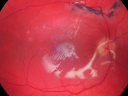
Giant Retinal Diaylsis Retinal Detachment - Post-op 2 months vision reasonable under silicone oil - Retina Attached579 views2 months post-op with silicone oil in eye.
|
|

X-linked Juvenille Retinoschisis - Peripheral Retinal Vascular Anomalies - Vitreous Hemorrhage - 8 Year Old Boy579 views8-year-old child OD 20/40, OS 20/50
OD: Vertical C/D ratio is 0.2. There are foveal cysts. There is also a retinal elevation inferiorly and there are patchy peripheral retinal hemorrhages.
OS: Vertical C/D ratio is 0.2. There are foveal cysts. There is peripheral retinal issues with some hemorrhage in some areas of peripheral retinoschisis.
|
|

579 views42-year-old man was seen in the office on October 5, 2011. He had noticed starting in August after a course of antibiotic and steroids, that he developed new spots in his vision in the right eye. He may have had an episode like this sometime in the past. He did take steroids a few years ago and his vision did change at that time, but then returned.
VISUAL ACUITY: OD 20/32, OS 20/32. The OCT scan of the right eye confirms subretinal fluid.
|
|

Serous Retinal Detachment - Hypertensive Retinopathy - 165/120 - Fluorescein Angiogram579 views48-year-old woman noticed when she was blinking there was a white flash in the left eye starting about a month ago. She sees it temporally. OD 20/16, OS 20/20. There is a serous retinal detachment superotemporal to the optic nerve falling short of the fovea. Her blood pressure 165/120.
|
|

Central Serous Retinopathy 25 year old professional baseball player - Atypical - Choroidal Hypo-perfusion?579 views
|
|

Proliferative Diabetic Retinopathy - Fresh and Old Scatter (Panretinal) Photocoagulation (Laser) - Neovascularization is regressing579 views1 month after initial laser
|
|

Proliferative Diabetic Retinopathy and Vitreous Hemorrhage - FA shows leakage of dye from NVE579 views
|
|

Choroidal-Melanoma with Retinal Hemorrhages579 viewsPatient comes in with blurry vision in his inferior visual field in his right eye. Fundus photography shows choroidal melanoma superiorly at 12-o'clock. Retinal hemorrhages scattered around the periphery.
|
|

Reticular Macular Disease (Dry Macular Degeneration) - Spectral Domain Optical Coherence Tomography Line Scan579 views85 year old woman with 20/25 vision OU and reticular macular disease
|
|
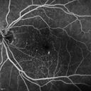
Cartwheel CNVM - Classic - wet AMD - evidence of possible old CSR - hydrophilic drusen stain early in FA579 views75 y.o Decreased Vision right eye for one year. VA OD: sc20/125+2 OS 20/25. Autofluorescence and SD OCT show evidence of old serous retinal detachment superior to the fovea suggesting a history of CSR. The macular lesion on FA shows a classic cartwheel appearance
|
|

BRAO and PAMM579 views
|
|

Posterior Uveal EffusionPosterior Uveal Effusion579 views
|
|

Cuticular Drusen - 26 year old female - Glomerulonephritis579 viewsVA 20/25 OU. Images are over 4 years
|
|

Multifocal Pattern Dystrophy579 views51 year old with normal vision - images look a lot like fundus flavimaculatus
|
|

Scleromalacia and Uveitis579 viewsPoor vision from uveitis and cataract. She was treated with Remicade and the uveitis abated and she had the right eye cataract done. The initial vision was 20/400 OD, 20/200 OS Final vision 20/60 OD, 20/32 OS
|
|

Choroidal Osteoma 13 Year Old579 views13 year old with 6 months of vision loss right eye and vision of 20/40
|
|

Sarcoidosis Multifocal Choroiditis579 views66 year old man with dense cataracts and recurrent uveitis. Images show multifocal choroidal granulomas from sarcoidosis more in the right eye than the left eye.
|
|

Punctate Inner Choroidopathy - New CNVM right eye579 views42 year old female with recent vision loss in the right eye - Right eye is 20/80 - Vision improved to 20/16 with Ranabizumab injections over 3 months
|
|

Metastatic Non Small Cell Lung Cancer to Choroid - Before External radiation579 views
|
|

Group 2a Juxtafoveal Retinal Telangiectasis (type 2)578 views58-year-old man decreased vision right eye and not better since cataract surgery one year ago. OD 20/40, OS 20/40.
|
|

Retinal Arterial Atheromata578 views68-year-old woman with records going back 7 years, which clearly document retinal arterial plaques in both eyes.
|
|

New BRVO Left Eye Old BRVO Right Eye - Left Eye Shows Twig Superior Vein Occlusion with Edema578 views90-year-old man decreased vision maybe one year since cataract surgery he is no longer able to read. OD 20/200, OS 20/200
|
|

Hypertensive Retinopathy One Year later with good Vision IgA Nephropathy578 viewsOD 20/30, OS 20/25 One year following initial episode.
|
|

Ruptured Retinal Arterial Macroaneurysm - Submacular Hemorrhage - Vision 20/400 never recovered - Hypertension 205/94578 views
|
|

Branch Retinal Arterial Occlusion 20/20 Vision - Symptoms for 1 Day578 views75-year-old man was seen in the office on 4/6/2010. He noticed a sudden vision change in the right eye yesterday morning. 20/20 Vision both Eyes
|
|

Ocular Ishemic Syndrome - Bilateral Carotid Occlusions - OD 20/60 OS 20/200 Enlarged Foveal Avascular Zone and Temporal Non-perfusion578 views75-year-old man had laryngeal cancer treated in 15 year ago with radiation. He recently was told that the carotid circulation is closed off on both sides. He does have a patent vertebral artery, which is supplying him pretty well and in addition to that his cardiac output is compromised by poor mitral valves. His consideration is to what can be done about that in the future.
VISUAL ACUITY: OD 20/60, OS 20/200
|
|

Chorioretinal Scar with Radiating Chorioretinal Folds578 views79-year-old woman has wet age-related macular degeneration in both eyes. The left eye had intravitreal injections of Avastin starting in May of 2009. Her vision remains good in the left eye and poor in the right eye.
VISUAL ACUITY: OD 8/200, OS 20/60.
|
|

Pigmented Choroidal Mass Right Eye - Melanoma vs. Nevus vs. Unilateral Melanocytic Proliferation - Initial Visit578 views71-year-old man is vision has been bothering him for about a month. He does not notice one eye particularly worse than the other. OD is 20/40, OS is 20/25 - Initial Visit
|
|

Chronic Pseudophakic Cystoid Macular Edema - Haptic Migrated into Anterior Chamber Through Peripheral Iridotomy578 views57-year-old man with congenital nystagmus. He had cataract surgery done 30 years ago and has decreased vision left eye for 6 months. OD is 20/50, OS is 20/100
|
|

Macular Branch Retinal Vein Occlusion and Adult Vitelliform Dystrophy578 views72-year-old woman 2 months post ECCE with poor vision since surgery especially left eye.
VISUAL ACUITY: OD 20/50, OS 20/200
|
|

Choroidal Melanoma (Medium) Exudative Retinal Detachment 578 views87-year-old man noticed a new onset floater a few months ago. OD is 20/40, OS is 20/30
|
|

Alphamethlyacyl COA Racemase Deficiency578 views
|
|

Serous Retinal Detachment - Hypertensive Retinopathy - 165/120 - Indocyanine Green Angiogram578 views48-year-old woman noticed when she was blinking there was a white flash in the left eye starting about a month ago. She sees it temporally. OD 20/16, OS 20/20. There is a serous retinal detachment superotemporal to the optic nerve falling short of the fovea. Her blood pressure 165/120.
|
|

Asymptomatic Juxtapapillary Choroidal Neovascular Membrane - Reticular Macular Disease (Wet AMD) 578 views
|
|

FA showing NVE, NVD, non- perfusion left eye inferonasally with neovascularization at the margin of the non-perfusion578 views
|
|

Chronic Central Serous Retinopathy - Fluorescein Angiogram578 views
|
|

Papilledema - Sagittal Cerebral Vein Thrombosis - Lung Transplant Cystic Fibrosis 36 Year Old Man578 viewsRNFL Analysis
|
|

Band Keratopathy/Neurotrophic Ulcer578 viewsPatient comes in with blind painful left eye. Slit lamp photos shows corneal diffuse scarring, descemets folds, corneal striae, band keratopathy, left eye. Proceed with Jupiter contact lens fitting on the left eye.
|
|

Polypoidal Choroidal Vasculopathy with Pulsating ICG polyp left eye578 views
|
|

Polypoidal Choroidal Vasculopathy with Pulsating ICG polyp left eye578 views
|
|
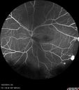
Sickle Retinopathy - Ultra Wide Field Angiogram - Heidelberg578 viewsLeakage from peripheral fronds of NV - HGB SC disease - 28 year old man with vision loss OS from acute vitreous hemorrhage
|
|

Choroidal Effusions and Serous retinal detachment with Systemic Testosterone and Cialis in 75 year old Man578 viewsB- Scan shows choroidal effusions in the left eye only
|
|

Posterior Uveal Effusion578 views
|
|

Hairy Cell Leukemia - Retinal Hemorrhage and twig Branch Vein Occlusion578 views79 year old man He has had hairy cell leukemia since 2002. He is in remission. His last blood tests were 9/2018. He just moved down here and needs a new leukemia doctor. His vision is fine.
VA OD: Dcc20/25
VA OS: Dcc20/25
IOP: TP: OD:12 OS:12
|
|

Confluent Soft Drusen with Subetinal Fluid - Right Eye578 viewsAsymptomatic 85 year old man. OCT shows pocket of subfoveal fluid in the center of the macula. This could be mechanical from the drusen preventing the retina from settling down. Testing did not reveal a CNVM and subsequent OCT 6 weeks later showed no change in structure.
|
|

Optic Nerve Pit Retinal Detachment - SD OCT of Nerve EDI shows Pit578 views9 Year old with morning glory disc who developed nerve pit RD. Raster scans of the optic nerve on EDI setting show the communication between the subretinal space and the fluid around the cerebrospinal fluid surrounding the optic nerve sheath.
|
|

Cystoid Macular Edema following Cataract surgery and vitrectomy for floaters578 views73 year old woman who had decreased vision 4 months following combined vitrectomy and cataract surgery. VA 20/63 improved in 1 month to 20/25 with ketorolac and prednisolone drops.
|
|
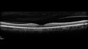
Posterior Scleritis - positive T-sign578 views62 year ols man was sick about 6 weeks ago and then 2 weeks ago the the left eye got red. He was treated for conjunctivitis and the eye did not get better. Topical steroids and oral non-steroidal anti-inflammatory medications did not help.
VA OD: Dcc20/25 NccJ5
VA OS: Dcc5'/200
The scleritis cleared within 2 weeks of starting high dose (80 mg) oral steroids. Blood tests were all negative.
|
|

Stellate non-hereditary idiopathic foveomacular retinoschisis (SNIFR) and optic nerve drusen578 views84 year old man No visual complaints. Not diabetic, cataract surgery 8 years ago. Meds: Omeprazole, Tamsulosin (Flomax)
VA 20/32 OD, 20/20 OS
|
|

Peripheral reticular pigment degeneration - pavingstone degeneration - age-related macular degeneration578 views60 year old healthy man with intermediate AMD and peripheral pigment degeneration and pavinstone degeneration. Vision is 20/20 OU and the patient is asymptomatic. No significant history of AMD and he is a non-smoker.
|
|

Wet AMD - Subfoveal CNVM - Excellent Response to Avastin577 views76-year-old woman two to three months of vision loss OD
Vision started at 20/200 and with Avastin improved to 20/25. She has required monthly injections and as of October 2010 has been treated about 16 times. Her left eye remains good at 20/20.
The OCT from post-treatment as well as pretreatment images are in this album.
|
|

PDR - Mild Vitreous Hemorrhage and Good Vision577 views58-year-old woman has diabetic retinopathy in both eyes with neovascularization of the optic nerve, worse in the right eye than the left eye. She has had panretinal laser in both eyes and vitreous hemorrhage in the left eye. Her vision, since I saw her last in June, is about the same.
VISUAL ACUITY: Vision OD is 20/25, OS is 20/20.
|
|
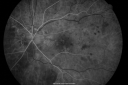
Diabetic Macular Edema with Circinate Exudate Early FA577 views74-year-old man has diabetic macular edema in both eyes and background diabetic retinopathy. His vision has blurred over the last few months. His blood sugars have been fluctuating.
VISUAL ACUITY: Vision OD is 20/25, OS is 20/80
|
|
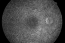
Multiple Evanescent White Dot Syndrome - Atypical Yellow Foveal Spot Right Eye577 views12-year-old had decreased vision in the left eye from reported amblyopia since around the year 2000. His right eye was doing fine until two days ago, when he had sudden severe vision loss. He sees a blurred spot in the middle of the vision in the right eye wherever he looks.
OD 20/60 with eccentric movement, OS 20/200. Pinhole 20/70. IOP: OD 14, OS 10.
|
|
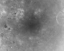
Clinically Significant Diabetic Macular Edema both Eyes with Exudates577 views56-year-old woman with diabetes for twelve years. She has noticed blurring of the vision in the right eye for about six months. OD is 20/70, OS is 20/25.
|
|
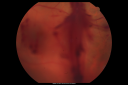
Diabetic Vitreous Hemorrhage Left Eye577 views35-year-old man diabetic for the last 25 years. His hemoglobin A1C has been running between 10 and 11. He was doing fine and then when he woke up from sleeping last night he noticed sudden vision loss in his left eye OD 20/30, OS 20/200.
|
|

Proliferative Diabetic Retinopathy in patient with Moyamoya Disease577 views28-year-old womandiabetic since age nine, and she also has had multiple strokes. OD is 20/30, OS is 20/30.
|
|

Central Retinal Artery Occlusion less than 24 hours - 69 year old man VA light perception577 views
|
|

577 views66-year-old woman had a gunshot wound to the head on August 12, 2010, with the entry wound just on the right side, just about below her temple, toward the lower part of the temple and the exit wound on the left side just above her cheek, a little behind the eye. Since then she recovered and did not need any surgery. When that all cleared she realized her vision was poor in both eyes. She has seen a neuro-ophthalmologist who thought she had traumatic optic neuropathy. OD light perception, OS 20/400.
|
|

Ocular Ishemic Syndrome - Bilateral Carotid Occlusions - OD 20/60 OS 20/200 Enlarged Foveal Avascular Zone and Temporal Non-perfusion577 views75-year-old man had laryngeal cancer treated in 15 year ago with radiation. He recently was told that the carotid circulation is closed off on both sides. He does have a patent vertebral artery, which is supplying him pretty well and in addition to that his cardiac output is compromised by poor mitral valves. His consideration is to what can be done about that in the future.
VISUAL ACUITY: OD 20/60, OS 20/200
|
|
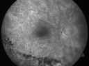
Macular Pucker 20/40 Vision - Pigment Bottom of Photo from Old Retinal Detachment 577 views59-year-old woman blurry vision in the right eye for about the last one or two years. She had cataract surgery in 1981, a retinal detachment repair with scleral buckle in 1988 in the right eye, and then she had lens implants in both eyes, anterior chamber lenses, in 1994. She did start taking Latisse for eyelashes about a year and a half ago.
VISUAL ACUITY: Vision OD is 20/40
(4 months later with a short course of antiinflammatory drops and new glasses her vision improved to 20/25)
|
|

Nanopthalmous (Microphthalmous) Chorioretinal Folds and Choroidal Thickening Both Eyes577 views67-year-old woman with microphthalmos/nanophthalmos Her vision has gradually been getting a little bit worse, especially in the right eye. Also the anterior chamber is getting crowded and there is concern about the need for cataract surgery. She has one sister and one cousin who also have nanophthalmos.
VISUAL ACUITY: OD 20/50, OS 20/80.
|
|

Myopic Macular Degeneration - Staphyloma - Recent Vision Loss Right Eye - Possible CNVM577 views83-year-old woman is a high myope with myopic macular degeneration. She had a retinal detachment repair in the right eye in 1968. I saw her in June of 2003. She had posterior colobomas and areas of pigment epithelial atrophy. She has noticed over the last six weeks substantial declining vision in the right eye. Prior to that, the vision in the two eyes was about the same. She uses Xalatan in the left eye and Alphagan in both eyes.
VISUAL ACUITY: Vision OD is 20/160, OS is 20/40
|
|
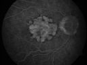
Geographic Atrophy 20/160 Vision577 views84-year-old woman has age-related macular degeneration in both eyes with foveal atrophy. She also has a central retinal vein occlusion
in the left eye for which she had intravitreal Kenalog and finally chorioretinal anastomosis laser a year
ago. VISUAL ACUITY: Vision OD is 20/160, OS is 7/200.
|
|

Acute Annular Outer Retinopathy for 3 Months - 25 YO Female FA577 views25-year-old female who, about three months ago, put on her sunglasses and noticed a smudge in the vision and at that point she noticed the right eye was hazy. She had a cold a week or two before the vision loss, but she didn’t have any fever. She has no focal neurological problems in her past history. VA 20/20 OU
|
|
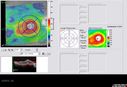
Post-Retinal Detachment Repair Occult Subfoveal CNVM577 viewsThis pleasant 56-year-old man has had a complex retinal detachment repair in the right eye. I most recently removed his silicone oil 3 months ago. His vision now is the best it has ever been.
VISUAL ACUITY: OD 4/200
|
|

Lumigan (Bimatoprost) Cystoid Macular Edema - Eye Better 1 month after Stopping Lumigan577 views 82-year-old man had cataract surgery in 1993. He has had glaucoma for seven or eight years for a long time. He is taking Lumigan only in the left eye. About a year ago he started in the right eye and then starting just about a few months ago he has noticed intermittent blurring in the vision in the right eye. Vision 20/25 OD, 20/20 OS
|
|

Lumigan (Bimatoprost) Cystoid Macular Edema - Eye Better 1 month after Stopping Lumigan577 views 82-year-old man had cataract surgery in 1993. He has had glaucoma for seven or eight years for a long time. He is taking Lumigan only in the left eye. About a year ago he started in the right eye and then starting just about a few months ago he has noticed intermittent blurring in the vision in the right eye. Vision 20/25 OD, 20/20 OS
|
|

Serous Retinal Detachment - Hypertensive Retinopathy - 165/120 577 views48-year-old woman noticed when she was blinking there was a white flash in the left eye starting about a month ago. She sees it temporally. OD 20/16, OS 20/20. There is a serous retinal detachment superotemporal to the optic nerve falling short of the fovea. Her blood pressure 165/120.
|
|

Serous Retinal Detachment - Hypertensive Retinopathy - 165/120 - Late - ICG shows leakage577 views48-year-old woman noticed when she was blinking there was a white flash in the left eye starting about a month ago. She sees it temporally. OD 20/16, OS 20/20. There is a serous retinal detachment superotemporal to the optic nerve falling short of the fovea. Her blood pressure 165/120.
|
|

Central Serous Retinopathy 25 year old professional baseball player - Atypical - Choroidal Hypo-perfusion?577 views
|
|
| 18117 files on 182 page(s) |
 |
|
|
|
|
56 | |
|
|
|
|


