|
|

Confluent Soft Drusen with Subetinal Fluid - Right Eye562 viewsAsymptomatic 85 year old man. OCT shows pocket of subfoveal fluid in the center of the macula. This could be mechanical from the drusen preventing the retina from settling down. Testing did not reveal a CNVM and subsequent OCT 6 weeks later showed no change in structure.
|
|

Choroidal Osteoma 13 Year Old562 views13 year old with 6 months of vision loss right eye and vision of 20/40
|
|

PDR - Mild Vitreous Hemorrhage and Good Vision561 views58-year-old woman has diabetic retinopathy in both eyes with neovascularization of the optic nerve, worse in the right eye than the left eye. She has had panretinal laser in both eyes and vitreous hemorrhage in the left eye. Her vision, since I saw her last in June, is about the same.
VISUAL ACUITY: Vision OD is 20/25, OS is 20/20.
|
|

Myopic CNVM - Wet - rx Avastin for 1 year561 views55-year-old woman OD is 20/40, OS is 20/20. There is a hyperpigmented disc diameter choroidal neovascular membrane with fluid touching the fovea.
|
|
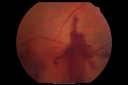
Diabetic Vitreous Hemorrhage Left Eye561 views35-year-old man diabetic for the last 25 years. His hemoglobin A1C has been running between 10 and 11. He was doing fine and then when he woke up from sleeping last night he noticed sudden vision loss in his left eye OD 20/30, OS 20/200.
|
|

Acute Zonal Occult Outer Retinopathy with Severe Peripheral Vision Loss Left Eye 561 views 58-year-old man developed a peripheral vision problem about 25 years ago. OD 20/30, OS 20/40.
|
|
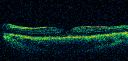
Perforating Steel Tip Dart Injury - 1 year later - no vitrectomy - 20/40 Vision 561 views16-year-old had a dart injury to the left eye one year ago. His vision is improving remarkably.
VISUAL ACUITY: OS: 20/40
|
|
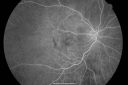
Best Disease for 30 Years - 20/50 Vision both eyes - 61 Year Old Woman561 views61-year-old woman was diagnosed with Best’s disease about 30 years ago. She and her many family members were tested, both at the VA Hospital and then at USF. OD 20/50, OS 20/50
|
|
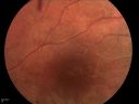
DUSN - Diffuse Unilateral Subacute Neuroretinitis - Nematode561 views
|
|
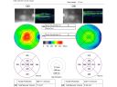
Macular Pucker 20/40 Vision 561 views59-year-old woman blurry vision in the right eye for about the last one or two years. She had cataract surgery in 1981, a retinal detachment repair with scleral buckle in 1988 in the right eye, and then she had lens implants in both eyes, anterior chamber lenses, in 1994. She did start taking Latisse for eyelashes about a year and a half ago.
VISUAL ACUITY: Vision OD is 20/40, OS is 20/20
(4 months later with a short course of antiinflammatory drops and new glasses her vision improved to 20/25)
|
|

Choroidal Melanoma - Exudative Retinal Detachment 82 Year Old Man 561 views82-year-old man who had 3 falls 2 weeks ago. After the falls he started checking his vision and noticed there was a veil over the left eye, which he had seen for about two weeks in the superior visual field. OD 20/25, OS 20/40
(patient was medically never well enough for brachytherapy and died 6 months later from heart disease)
|
|

Pigmented Choroidal Mass Right Eye - Melanoma vs. Nevus vs. Unilateral Melanocytic Proliferation - 4 Month Follow-Up Images561 views71-year-old man his vision 4 month follow-up. Vision is 20/60 and patient sees flashing lights. Lesion has grown slightly in the superior half of the macula.
|
|
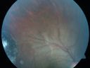
Congenital Toxoplasmosis with non-macular Scars Both Eyes - Recent Reactivation (recurrence) Right Eye561 views34-year-old man has congenital toxoplasmosis with four week history of foggy vision and new floaters in the right eye. OD is 20/40, OS 20/30
|
|

Choroidal Melanoma (Medium) Exudative Retinal Detachment 561 views87-year-old man noticed a new onset floater a few months ago. OD is 20/40, OS is 20/30
|
|

Acute Central Serous Retinopathy - CSR - Steroid Induced (probably) - Good Vision 561 views42-year-old man was seen in the office on October 5, 2011. He had noticed starting in August after a course of antibiotic and steroids, that he developed new spots in his vision in the right eye. He may have had an episode like this sometime in the past. He did take steroids a few years ago and his vision did change at that time, but then returned.
VISUAL ACUITY: OD 20/32, OS 20/32. The OCT scan of the right eye confirms subretinal fluid.
|
|

Lumigan (Bimatoprost) Cystoid Macular Edema - Eye Better 1 month after Stopping Lumigan561 views 82-year-old man had cataract surgery in 1993. He has had glaucoma for seven or eight years for a long time. He is taking Lumigan only in the left eye. About a year ago he started in the right eye and then starting just about a few months ago he has noticed intermittent blurring in the vision in the right eye. Vision 20/25 OD, 20/20 OS
|
|

Lumigan (Bimatoprost) Cystoid Macular Edema - Eye Better 1 month after Stopping Lumigan561 views 82-year-old man had cataract surgery in 1993. He has had glaucoma for seven or eight years for a long time. He is taking Lumigan only in the left eye. About a year ago he started in the right eye and then starting just about a few months ago he has noticed intermittent blurring in the vision in the right eye. Vision 20/25 OD, 20/20 OS
|
|

Serous Retinal Detachment - Hypertensive Retinopathy - 165/120 - Indocyanine Green Angiogram561 views48-year-old woman noticed when she was blinking there was a white flash in the left eye starting about a month ago. She sees it temporally. OD 20/16, OS 20/20. There is a serous retinal detachment superotemporal to the optic nerve falling short of the fovea. Her blood pressure 165/120.
|
|

Non-specific (uncharaterized / unknown) Macular Dystrophy 561 views59-year-old man has macular dystrophy in both eyes. He had some vision changes in medical school in the 1980s and saw Dr. Gass for that. He had some pigment epithelial irregularities in both eyes. He had strabismus as a child and had muscle surgery. He is color blind, along with many people in his family, and as far as he knows, it is congenital.
20/25, 20/30
|
|

Pigmented Peripheral Retinal Degeneration561 views42-year old male comes in for routine eye exam and to follow up on peripheral retinal degeneration in both eyes. VA is 20/20, right eye and 20/25, left eye. Patient is asymptomatic with no visual complaints.
|
|

Cartwheel CNVM - Classic - wet AMD - evidence of possible old CSR561 views75 y.o Decreased Vision right eye for one year. VA OD: sc20/125+2 OS 20/25. Autofluorescence and SD OCT show evidence of old serous retinal detachment superior to the fovea suggesting a history of CSR. The macular lesion on FA shows a classic cartwheel appearance
|
|

Vitreomacular traction - recent vision loss Right Eye - initially worse after Jetrea561 views
|
|

Unilateral Acute Idiopathic Maculopathy (UAIM)561 views41 year old with serous retinal detachment OS and sudden vision loss. He had a viral prodrome. Coxscackie IgG was high but no acute infection was documented. Vision improved in about 2 months without therapy. - Right eye has spot nasal to the optic nerve
|
|

Presumed Ocular Histoplasmosis with new CNVM in right and better eye - VA 20/80 OD; 20/800 OS561 views
|
|

Central retinal artery and vein occlusion - Protein C Deficiency 33 year old male561 views33 year old male with vision loss for 3 hours. VA 20/12 right eye, 5/200 left eye. He has a protein C deficiency. The left eye recovered vision in about 1 month to 20/16. FA shows very slow recirculation time.
|
|

Recurrent MEWDS Left Eye - Teenage Girl561 viewsPatient presented in 2014 (first images) 2 months following vision loss left eye. Vision went from 20/60 - 20/20. OCT showed outer retinal irregularities which normalized. Then 3 years later in 2017 she had full blown MEWDS. She presented acutely and all the images are typical. Both episodes where in the same time of year, about 3 months after her flu shot for sports.
|
|

Prefoveal Opercula and early Lamellar Macular Hole561 views73 year old man with 20/32 vision and pre-foveal opercula. The OCT shows very early lamellar hole with lamellar hole epiretinal proliferation
|
|

Sarcoidosis Multifocal Choroiditis561 views66 year old man with dense cataracts and recurrent uveitis. Images show multifocal choroidal granulomas from sarcoidosis more in the right eye than the left eye.
|
|

Hemangioma OS with good vison - 01-29-18561 viewsImages 7 years following initial uploaded images
|
|

BDR and Diabetic Macular Edema with Fresh Laser Right Eye560 views60-year old man OD 20/60, OS 20/50 worsening vision both eyes
|
|
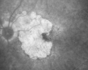
Geographic Atrophy and Age-Related Macular Degeneration - Choriocapillaris is Largely Still Intact560 views85-year-old woman has age-related macular degeneration in both eyes with geographic atrophy. She has noticed her vision getting slightly worse in the past 6 months. VISUAL ACUITY: OD 20/70, OS 20/400
|
|

Juxtfoveal Telangiectasis Group 2a - Type II - MacTel - also Juxtapapillary Asymptomatic CNVM Right Eye560 views82-year-old woman who has juxtafoveal retinal telangiectasis in both eyes. She recently had cataract surgery in the right eye and the vision in the right eye is still limited. OD 20/100, OS 20/40.
|
|

560 views
|
|

Central Retinal Artery Occlusion less than 24 hours - 69 year old man VA light perception560 views
|
|

Macular Pucker 20/40 vision and recent cataract surgery560 views 73-year-old man distortion and vision loss since ECCE 6 months ago OS. Patient taking anti-inflammatory drops. VISUAL ACUITY: OD 20/30, OS 20/40.
|
|

Myopic Macular Degeneration - Staphyloma - Recent Vision Loss Right Eye - Possible CNVM560 views83-year-old woman is a high myope with myopic macular degeneration. She had a retinal detachment repair in the right eye in 1968. I saw her in June of 2003. She had posterior colobomas and areas of pigment epithelial atrophy. She has noticed over the last six weeks substantial declining vision in the right eye. Prior to that, the vision in the two eyes was about the same. She uses Xalatan in the left eye and Alphagan in both eyes.
VISUAL ACUITY: Vision OD is 20/160, OS is 20/40
|
|

Acute Annular Outer Retinopathy for 3 Months - 25 YO Female FA560 views25-year-old female who, about three months ago, put on her sunglasses and noticed a smudge in the vision and at that point she noticed the right eye was hazy. She had a cold a week or two before the vision loss, but she didn’t have any fever. She has no focal neurological problems in her past history. VA 20/20 OU
|
|

Acute Annular Outer Retinopathy for 3 Months - 25 YO Female Fellow Normal Eye560 views25-year-old female who, about three months ago, put on her sunglasses and noticed a smudge in the vision and at that point she noticed the right eye was hazy. She had a cold a week or two before the vision loss, but she didn’t have any fever. She has no focal neurological problems in her past history. VA 20/20 OU
|
|

Pigmented Choroidal Mass Right Eye - Melanoma vs. Nevus vs. Unilateral Melanocytic Proliferation - Initial Visit560 views71-year-old man is vision has been bothering him for about a month. He does not notice one eye particularly worse than the other. OD is 20/40, OS is 20/25 - Initial Visit
|
|

Pigmented Choroidal Mass Right Eye - Melanoma vs. Nevus vs. Unilateral Melanocytic Proliferation - Initial Visit560 views71-year-old man is vision has been bothering him for about a month. He does not notice one eye particularly worse than the other. OD is 20/40, OS is 20/25 - Initial Visit71-year-old man is vision has been bothering him for about a month. He does not notice one eye particularly worse than the other. OD is 20/40, OS is 20/25 - Initial Visit
|
|

Macular Branch Retinal Vein Occlusion and Adult Vitelliform Dystrophy560 views72-year-old woman 2 months post ECCE with poor vision since surgery especially left eye.
VISUAL ACUITY: OD 20/50, OS 20/200
|
|

Cuticular Drusen (Basal Laminar Drusen) and Bilateral Chronic Subfoveal Fluid (Serous Retinal Detachment) unresponsive to monthly Avastin Very Early ICG OD560 views66-year-old man with bilateral subfoveal fluid unresponsive to one year of monthly avastin therapy.
OD 20/160, Pinhole 20/80. OS 20/63, Pinhole 20/50. OCT scan shows subretinal fluid with normal choroidal thickness. Fluorescein angiogram shows cuticular/basal laminar drusen. ICG shows no foci of choriocapillaris leakage.
|
|

Lumigan (Bimatoprost) Cystoid Macular Edema - Eye Better 1 month after Stopping Lumigan560 views 82-year-old man had cataract surgery in 1993. He has had glaucoma for seven or eight years for a long time. He is taking Lumigan only in the left eye. About a year ago he started in the right eye and then starting just about a few months ago he has noticed intermittent blurring in the vision in the right eye. Vision 20/25 OD, 20/20 OS
|
|

Retinitis Pigmentosa Sine Pigmento or Unknown Dystrophy - Peripheral Superficial White Spots and Recurrent CME560 views1 month of treatment with diamox 500 mg BID- cme is a little better
|
|
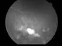
Uveitis and Retinal Whitening - 04-27-12 - Prior to PPV OD - Possible Primary Ocular Lymphoma 560 views
|
|
Primary Ocular Lymphoma - 051812 560 views
|
|
Primary Ocular Lymphoma - 051812 560 views
|
|

Primary Ocular Lymphoma - 020513 - FUndus Autofluorescence560 views
|
|

Medium Choroidal Melanoma Left Eye 560 views
|
|

Reticular Macular Disease (Pseudo-drusen) Both Eyes - Occult CNVM and Macular Edema Right Eye 560 viewsInfra-Red
|
|

560 views
|
|

BDR FAF560 views
|
|

Proliferative Diabetic Retinopathy and Vitreous Hemorrhage - FA shows leakage of dye from NVE560 views
|
|

Chronic Central Serous Retinopathy - Fluorescein Angiogram560 views
|
|

560 views
|
|

Sickle Retinpoathy SC Disease - Vitreous Hemorrhage Left Eye - 29 Year old560 viewsFrond of NV in the periphery
|
|

Posterior Uveal Effusion560 views
|
|

Macular Edema and dry AMD - excellent example of why FA and ICG are necessary in management of wet AMD560 viewsThis 72 year old woman has an OCT that looks like wet AMD. Only her FA and ICG show that she has inflammatory macular edema. She had no clinical signs or symptoms of uveitis. Nevertheless, the macular edema responded to subtenon's kenalog.
|
|

Syphilitic Chorioretinitis560 viewsSevere outer retinal atrophy and RPE non-geographic atrophy from syphilis. Vision improved from 20/63 to 20/32 with therapy. Images look a lot like DUSN. Patient had several surgeries (vitrectomy for floaters, vitrectomy for macular pucker, Avastin injections and macular laser for edema) prior by other retina specialists prior to being correctly diagnosed and treated.
|
|

Syphilitic Chorioretinitis560 viewsSevere outer retinal atrophy and RPE non-geographic atrophy from syphilis. Vision improved from 20/63 to 20/32 with therapy. Images look a lot like DUSN. Patient had several surgeries (vitrectomy for floaters, vitrectomy for macular pucker, Avastin injections and macular laser for edema) prior by other retina specialists prior to being correctly diagnosed and treated.
|
|

Asymptomatic wet AMD - Subfoveal CNM both eyes560 views82 year old woman - 20/25 OU. Images show occult subfoveal CNVM both eyes best seen on late ICG. The left eye also has probable polypoidal choroidal vasculopathy. No treatment was recommended on the first visit - follow-up at 1 month.
|
|

Scleromalacia and Uveitis560 viewsPoor vision from uveitis and cataract. She was treated with Remicade and the uveitis abated and she had the right eye cataract done. The initial vision was 20/400 OD, 20/200 OS Final vision 20/60 OD, 20/32 OS
|
|

Scleromalacia and Uveitis560 viewsPoor vision from uveitis and cataract. She was treated with Remicade and the uveitis abated and she had the right eye cataract done. The initial vision was 20/400 OD, 20/200 OS Final vision 20/60 OD, 20/32 OS
|
|

Choroidal Nevus - Macula - Flat560 views37 year old female with 5 year stable nevus Left eye - OCT shows flat nevus (EDI scan)
|
|

Punctate Inner Choroidopathy - New CNVM right eye560 views42 year old female with recent vision loss in the right eye - Right eye is 20/80 - Vision improved to 20/16 with Ranabizumab injections over 3 months
|
|

Cat Scratch - Neuroretinitis OD AND retinitis OS - Bartonella henselae positive560 views27 year old female with exposure to cats and prior fever. Reports with recent vision loss right eye. She was treated with doxycycline and within 2 weeks the right eye looks better. Then she skipped all follow-up visits. The right eye presented 20/200 and the left 20/16
|
|

Grade 4 hypertensive retinopathy560 views55 year old man Hhe did have headaches a few weeks ago and also some nausea and vomiting. He noticed for a few weeks the vision in the left eye was blurred and that the vision in the left eye is dark.
VA OD: Dcc20/20
VA OS: Dcc20/100-1
He was admitted directly to ICU for blood pressure control (290 / 170 mmHg)
3 weeks later his VA increased to 20/20 OD, 20/32 OS
|
|

Stellate non-hereditary idiopathic foveomacular retinoschisis (SNIFR) and optic nerve drusen560 views84 year old man No visual complaints. Not diabetic, cataract surgery 8 years ago. Meds: Omeprazole, Tamsulosin (Flomax)
VA 20/32 OD, 20/20 OS
|
|

Stellate non-hereditary idiopathic foveomacular retinoschisis (SNIFR) and optic nerve drusen560 views84 year old man No visual complaints. Not diabetic, cataract surgery 8 years ago. Meds: Omeprazole, Tamsulosin (Flomax)
VA 20/32 OD, 20/20 OS
|
|

Myopic CNVM - Wet - rx Avastin for 1 year559 views55-year-old woman OD is 20/40, OS is 20/20. There is a hyperpigmented disc diameter choroidal neovascular membrane with fluid touching the fovea.
|
|
Macular Scar Left Eye from Age-Related Macular Degeneration559 views81-year-old woman has a macular scar in the left eye and then unfortunately developed a central retinal artery occlusion in the right eye OD 20/100, OS 6/200.
|
|

Cytomegalovirus Retinitis - Partially Treated and AIDS retinopathy559 views44-year-old man a month agoe was diagnosed with HIV and he had pneumonia. He has had blurred vision for about a month, more in the left eye than the right eye. He has not yet seen anyone to start anti-AIDS therapy. He was given a medical doctor's name for a follow-up visit. He thinks his CD4 count is somewhere in the low 100s. Vision OU is 20/30
|
|
Cotton Wool Spots Both Eyes after Strep Pneumonia and Cardiac ICU Stay 1 Month Ago 559 views60-year-old man was sick in the hospital in 1 month ago with streptococcal pneumonia. He was in the cardiac intensive care unit and at one point he was in critical condition. He has no visual complaints. OD 20/20, OS 20/25.
|
|

Chronic Central Serous Retinopathy treated with Photodynamic Laser and Visudyne559 views48-year-old man has had waxing and waning vision in the left eye for the last year from central serous retinopathy. OD is 20/16, OS is 20/30.
|
|

559 views
|
|
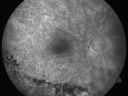
Macular Pucker 20/40 Vision - Pigment Bottom of Photo from Old Retinal Detachment 559 views59-year-old woman blurry vision in the right eye for about the last one or two years. She had cataract surgery in 1981, a retinal detachment repair with scleral buckle in 1988 in the right eye, and then she had lens implants in both eyes, anterior chamber lenses, in 1994. She did start taking Latisse for eyelashes about a year and a half ago.
VISUAL ACUITY: Vision OD is 20/40
(4 months later with a short course of antiinflammatory drops and new glasses her vision improved to 20/25)
|
|

Nanopthalmous (Microphthalmous) Chorioretinal Folds and Choroidal Thickening Both Eyes559 views67-year-old woman with microphthalmos/nanophthalmos Her vision has gradually been getting a little bit worse, especially in the right eye. Also the anterior chamber is getting crowded and there is concern about the need for cataract surgery. She has one sister and one cousin who also have nanophthalmos.
VISUAL ACUITY: OD 20/50, OS 20/80.
|
|

Diabetic Retinopathy - Foveal Laser Scar Right Eye559 views74-year-old woman has diabetic retinopathy in both eyes. She has had focal laser in her right eye done elsewhere in the mid 90’s and unfortunately the laser was fairly close to the center and as time progressed she developed foveal atrophy around the scars. She has noticed her vision declining since she was here last a year ago. She does take Travatan in both eyes at night for glaucoma.
VISUAL ACUITY: OD 20/200, OS 20/40.
|
|

Myopic Traction Maculopathy - Vitreoretinal Traction - Possible Occult Neovascular Membrane559 views58-year-old woman was seen in the office on June 30, 2011. In 2002, her vision was 20/30. Subsequent to that she had her left eye made her distance eye, which was dominate. She had LASIK surgery on the right eye to make that her near eye and she was getting along well until about a year ago when she noticed substantial vision loss in the left eye only. VISUAL ACUITY: The near eye is 20/200, but she reads J2. OS 20/100- and J16.
|
|

New Subfoveal Classic CNVM OS - Wet AMD - Macular Scar Congenital Toxoplasmosis Right Eye 559 views77-year-old woman as had poor vision in the right eye since birth from congenital toxoplasmosis of her macula in the right eye. Two to three weeks ago when she noticed vision loss in the left eye. VISUAL ACUITY: OD 2/200, OS 20/200.
|
|
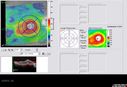
Post-Retinal Detachment Repair Occult Subfoveal CNVM559 viewsThis pleasant 56-year-old man has had a complex retinal detachment repair in the right eye. I most recently removed his silicone oil 3 months ago. His vision now is the best it has ever been.
VISUAL ACUITY: OD 4/200
|
|

Serous Retinal Detachment - Hypertensive Retinopathy - 165/120 - Late - ICG shows leakage559 views48-year-old woman noticed when she was blinking there was a white flash in the left eye starting about a month ago. She sees it temporally. OD 20/16, OS 20/20. There is a serous retinal detachment superotemporal to the optic nerve falling short of the fovea. Her blood pressure 165/120.
|
|
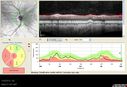
Optic Nerve Hypoplasia - 43 Year Old Man - declining vision559 views43-year-old man was diagnosed with optic nerve hypoplasia at age fifteen at the National Institute of Health. His vision seems to be declining lately.
Vision OD is 20/80, OS is 20/120
|
|

Asymptomatic Juxtapapillary Choroidal Neovascular Membrane - Reticular Macular Disease (Wet AMD) 559 views
|
|

Pigmented Peripheral Retinal Degeneration559 views42-year old male comes in for routine eye exam and to follow up on peripheral retinal degeneration in both eyes. VA is 20/20, right eye and 20/25, left eye. Patient is asymptomatic with no visual complaints.
|
|

Proliferative Diabetic Retinopathy - Fresh and Old Scatter (Panretinal) Photocoagulation (Laser) - Neovascularization is regressing559 views1 month after initial laser
|
|

Vitreomacular traction - recent vision loss left eye - FA559 views
|
|

Branch Retinal Vein Occlusion with worse macular edema from hypertension - FA shows Edema Right Eye559 views
|
|

Stasis Retinopathy, Central Retinal Vein Occlusion559 viewsYoung male who complains of decreased vision in the left eye. VA is 20/20 in the left eye. Fundus photo shows mild central retinal vein occlusion in the left eye.
|
|

Iris Nevus559 viewsPatient came in for cataract evaluation. VA 20/200, right eye, 20/60, left eye. Slit lamp photography shows iris nevus 3-4 mm located nasally at 9-o'clock in the left eye.
|
|

Bilateral Papilledema 559 viewsYoung male presents with decreased vision in both eyes. Fundus photos reveal swollen optic nerves in both eyes.
|
|

Retinitis Pigmentosa Sine Pigmento 559 views
|
|
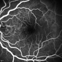
Polypoidal Choroidal Vasculopathy with Pulsating ICG polyp left eye559 views
|
|

Hairy Cell Leukemia - Retinal Hemorrhage and twig Branch Vein Occlusion559 views79 year old man He has had hairy cell leukemia since 2002. He is in remission. His last blood tests were 9/2018. He just moved down here and needs a new leukemia doctor. His vision is fine.
VA OD: Dcc20/25
VA OS: Dcc20/25
IOP: TP: OD:12 OS:12
|
|

Macular Edema and dry AMD - excellent example of why FA and ICG are necessary in management of wet AMD559 viewsThis 72 year old woman has an OCT that looks like wet AMD. Only her FA and ICG show that she has inflammatory macular edema. She had no clinical signs or symptoms of uveitis. Nevertheless, the macular edema responded to subtenon's kenalog.
|
|

Syphilitic Chorioretinitis559 viewsSevere outer retinal atrophy and RPE non-geographic atrophy from syphilis. Vision improved from 20/63 to 20/32 with therapy. Images look a lot like DUSN. Patient had several surgeries (vitrectomy for floaters, vitrectomy for macular pucker, Avastin injections and macular laser for edema) prior by other retina specialists prior to being correctly diagnosed and treated.
|
|

Ocular Lymphoma - non-Hodgkins - B-Cell559 viewsMultifocal sub-RPE lesions in the left eye of a patient with history of Non-hodgkins B-cell Lymphoma last active 2 years ago
|
|

Scleromalacia and Uveitis559 viewsPoor vision from uveitis and cataract. She was treated with Remicade and the uveitis abated and she had the right eye cataract done. The initial vision was 20/400 OD, 20/200 OS Final vision 20/60 OD, 20/32 OS
|
|

Autoimmune Retinopathy559 views91 year old female with severe vision loss over the past 3 years. She has shimmery scotomas. The main findings are outer retinal atrophy evident on OCT and FAF. VA 20/80 OD and 20/125 OS
|
|

Subtle Branch Retinal Artery Occlusion559 views68 year old man with shadow in the left eye yesterday. IR and OCT show subtle retinal swelling. FA is normal
|
|
| 18004 files on 181 page(s) |
 |
|
|
|
|
57 | |
|
|
|
|


