|
|
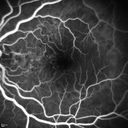
Polypoidal Choroidal Vasculopathy with Pulsating ICG polyp left eye558 views
|
|

Choroideremia - Female Carrier - Patchy Atrophy of Choroid and RPE - Lyonization558 views
|
|

Acute Posterior Multifocal Placoid Pigment Epitheliopathy - 4 years after onset558 views4 years after presentation - vision 20/80 OD 20/16 OS and there is subretinal fibrosis
|
|

Macular Edema and dry AMD - excellent example of why FA and ICG are necessary in management of wet AMD558 viewsThis 72 year old woman has an OCT that looks like wet AMD. Only her FA and ICG show that she has inflammatory macular edema. She had no clinical signs or symptoms of uveitis. Nevertheless, the macular edema responded to subtenon's kenalog.
|
|

Optic Nerve Pit Retinal Detachment - SD OCT of Nerve EDI shows Pit558 views9 Year old with morning glory disc who developed nerve pit RD. Raster scans of the optic nerve on EDI setting show the communication between the subretinal space and the fluid around the cerebrospinal fluid surrounding the optic nerve sheath.
|
|

CRVO with Paracentral Acute Middle Maculopath - PAMM558 views56 year old woman with sudden vision loss, hemorrhages in all four quadrants. FA shows good retinal circulation. The OCT shows PAMM lesions in the affected eye. Vision did improve on second visit from 20/160 to 20/60 in about a month
|
|

Scleromalacia and Uveitis558 viewsPoor vision from uveitis and cataract. She was treated with Remicade and the uveitis abated and she had the right eye cataract done. The initial vision was 20/400 OD, 20/200 OS Final vision 20/60 OD, 20/32 OS
|
|

Scleromalacia and Uveitis558 viewsPoor vision from uveitis and cataract. She was treated with Remicade and the uveitis abated and she had the right eye cataract done. The initial vision was 20/400 OD, 20/200 OS Final vision 20/60 OD, 20/32 OS
|
|
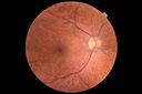
Hemangioma OS with good vison - 01-29-18558 viewsImages 7 years following initial uploaded images
|
|

Punctate Inner Choroidopathy - New CNVM right eye558 views42 year old female with recent vision loss in the right eye - Right eye is 20/80 - Vision improved to 20/16 with Ranabizumab injections over 3 months
|
|

Autoimmune Retinopathy558 views91 year old female with severe vision loss over the past 3 years. She has shimmery scotomas. The main findings are outer retinal atrophy evident on OCT and FAF. VA 20/80 OD and 20/125 OS
|
|
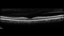
Posterior Scleritis - positive T-sign558 views62 year ols man was sick about 6 weeks ago and then 2 weeks ago the the left eye got red. He was treated for conjunctivitis and the eye did not get better. Topical steroids and oral non-steroidal anti-inflammatory medications did not help.
VA OD: Dcc20/25 NccJ5
VA OS: Dcc5'/200
The scleritis cleared within 2 weeks of starting high dose (80 mg) oral steroids. Blood tests were all negative.
|
|

Subtle Branch Retinal Artery Occlusion558 views68 year old man with shadow in the left eye yesterday. IR and OCT show subtle retinal swelling. FA is normal
|
|

PDR - Macula Line Scan557 views58-year-old woman has diabetic retinopathy in both eyes with neovascularization of the optic nerve, worse in the right eye than the left eye. She has had panretinal laser in both eyes and vitreous hemorrhage in the left eye. Her vision, since I saw her last in June, is about the same.
VISUAL ACUITY: Vision OD is 20/25, OS is 20/20.
|
|

Macular Pucker in eye following giant retinal tear RD repair pre-op OCT557 viewsnow that the bubble is gone, but he is starting to notice distortion of central vision. He had a giant retinal tear retinal detachment repair emergently done on November 8, 2007, and subsequent laser on November 15th.
VISUAL ACUITY: Vision OD 20/400.
6 weeks post-op VA 20/40
|
|

Myopic CNVM - Wet - rx Avastin for 1 year557 views55-year-old woman OD is 20/40, OS is 20/20. There is a hyperpigmented disc diameter choroidal neovascular membrane with fluid touching the fovea.
|
|

Diabetic Patient with Macular Edema and Blood Pressure 200/95557 views49-year-old decreasing vision over the last year. OD is 20/80, OS 20/80. blood pressure which was 200/95.
|
|

Purtchers Retinopathy with Bilateral Central Retinal Artery Occlusions 557 views50-year-old man traumatic chest compression and leg crushing injury. OD 1/200, OS 3/200
|
|

Chronic Pseudophakic Cystoid Macular Edema in a Patient with Diabetes557 views74-year-old woman has chronic cystoid macular edema in the left eye. She has had vision loss intermittently in that eye for 2 years, the right eye, unfortunately, lost vision from a central retinal vein occlusion in 6 years ago.
|
|

Central Retinal Artery Occlusion less than 24 hours - 69 year old man VA light perception557 views
|
|

Branch Retinal Arterial Occlusion 20/20 Vision - Symptoms for 1 Day557 views75-year-old man was seen in the office on 4/6/2010. He noticed a sudden vision change in the right eye yesterday morning. 20/20 Vision both Eyes
|
|

557 views66-year-old woman had a gunshot wound to the head on August 12, 2010, with the entry wound just on the right side, just about below her temple, toward the lower part of the temple and the exit wound on the left side just above her cheek, a little behind the eye. Since then she recovered and did not need any surgery. When that all cleared she realized her vision was poor in both eyes. She has seen a neuro-ophthalmologist who thought she had traumatic optic neuropathy. OD light perception, OS 20/400.
|
|

Birdshot Chorioretinitis - Retinal Vasculitis - HLA A29 Positive 557 views64-year-old woman three or four year’s fluctuations in her vision. She mostly notices them in bright light her vision glares out and she has difficulty seeing. PMHx: malaria in 1948 that was treated. She was told at one point she could possibly have had Familial Mediterranean fever.
She does intermittently get rashes around her arms. She has arthritis in her knees and ankles.
VISUAL ACUITY: OD 20/40, OS 20/50 - HLA - A29 Positive
|
|

Choriodal Neovascular Membrane - Subfoveal - Pseudo-Retinitis Pigmentosa - 50 years post Retinal Detachment Repair557 views86-year-old with a history of decreased vision in the right eye. The patient reports she has lost vision in the right eye over the past two months or so. She reports the vision was always compromised in that eye since retinal detachment surgery 50 years ago with a scleral buckler. Vision dropped from 20/50 to 20/200 right eye in 2 months.
|
|

Retinal Detachment with Subretinal Fibrosis - Chronic with new Foveal Involvement 25 Year Old557 views25-year-old man vision loss in the left eye for a month. He noticed a shadow up above, which went through his central vision. He has not had any injuries in the left eye.
VISUAL ACUITY: Vision OD is 20/20, OS is 20/400
|
|
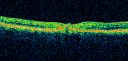
Juxtafoveal Telangiectasis - After Hemorrhage Absorbed Right Eye with Avastin Injections557 views73-year-old woman has juxtafoveal retinal telangiectasis in both eyes. The right eye has stage 5 disease. Her last Avastin treatment in the right eye was in 6 months ago. The right eye has been the worst of her two eyes for years.
VISUAL ACUITY: Vision OD is 20/200, OS is 20/50.
|
|

Macular Pucker - 1 year after Vitrectomy - 20/25 Vision - Postoperative Images557 views60-year-old man had vitrectomy for a macular pucker in the right eye a year ago. His vision is good.
VISUAL ACUITY: Vision OD is 20/25 Eye is now pseudophakic
|
|

Geographic Atrophy 20/160 Vision557 views84-year-old woman has age-related macular degeneration in both eyes with foveal atrophy. She also has a central retinal vein occlusion
in the left eye for which she had intravitreal Kenalog and finally chorioretinal anastomosis laser a year
ago. VISUAL ACUITY: Vision OD is 20/160, OS is 7/200.
|
|

AIDS - HIV Retinopathy - Cotton Wool Spots557 views54-year-old woman HIV positive for 18 years. Her CD4 count is currently less than 50 and she was hospitalized for pneumocystis carinii pneumonia 5 months ago. She has noticed just for the last few weeks a few blind spots in the paracentral vision. Predominantly out of her right eye when she closes her left eye, she sees a few temporal blind spots. VA 20/25 in both eyes.
|
|

Acute Annular Outer Retinopathy for 3 Months - 25 YO Female Fellow Normal Eye557 views25-year-old female who, about three months ago, put on her sunglasses and noticed a smudge in the vision and at that point she noticed the right eye was hazy. She had a cold a week or two before the vision loss, but she didn’t have any fever. She has no focal neurological problems in her past history. VA 20/20 OU
|
|

Choroidal Hemangioma557 views
|
|

Retinitis Pigmentosa Newly Diagnosed 55 Year Old557 views55-year-old noticed his vision declining recently, over the last few weeks, particularly in the right eye. He said previous to that the vision was not normal but was pretty good. OD is 20/100, OS is 20/50
OCT SCAN: There is macular thickening in both eyes with intraretinal cysts.
|
|
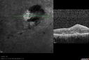
Post-Retinal Detachment Repair Occult Subfoveal CNVM557 viewsThis pleasant 56-year-old man has had a complex retinal detachment repair in the right eye. I most recently removed his silicone oil 3 months ago. His vision now is the best it has ever been.
VISUAL ACUITY: OD 4/200
|
|

Serous Retinal Detachment - Hypertensive Retinopathy - 165/120 - Fluorescein Angiogram557 views48-year-old woman noticed when she was blinking there was a white flash in the left eye starting about a month ago. She sees it temporally. OD 20/16, OS 20/20. There is a serous retinal detachment superotemporal to the optic nerve falling short of the fovea. Her blood pressure 165/120.
|
|

FA showing NVE, NVD, non- perfusion left eye inferonasally with neovascularization at the margin of the non-perfusion557 views
|
|

Reticular Macular Disease (Pseudo-drusen) Both Eyes - Occult CNVM and Macular Edema Right Eye 557 viewsInfra-Red
|
|

Reticular Macular Disease (Pseudo-drusen) Both Eyes - Occult CNVM and Macular Edema Right Eye 557 viewsIndocyanine Green Angiogram
|
|

Branch Retinal Vein Occlusion with worse macular edema from hypertension - FA shows Edema Right Eye557 views
|
|

Choroidal Effusions and Serous retinal detachment with Systemic Testosterone and Cialis in 75 year old Man557 viewsB- Scan shows choroidal effusions in the left eye only
|
|

Retinitis Pigmentosa - Autosomal Recessive - 82 year old man with Good Vision557 viewsVA 20/25, 20/32 - Bone spicules in periphery - FAF is very symmetric
|
|

Posterior Uveal Effusion557 views
|
|

Presumed Ocular Histoplasmosis with new CNVM in right and better eye - VA 20/80 OD; 20/800 OS557 views
|
|

Stargardt's Macular Dystrophy ABCA4 positive557 views Fundus Autofluorescence shows central atrophy with Hyper FAF pisciform triradiate lesions. Images show some progression over 3 years of macular dystrophy.
|
|

Hairy Cell Leukemia - Retinal Hemorrhage and twig Branch Vein Occlusion557 views79 year old man He has had hairy cell leukemia since 2002. He is in remission. His last blood tests were 9/2018. He just moved down here and needs a new leukemia doctor. His vision is fine.
VA OD: Dcc20/25
VA OS: Dcc20/25
IOP: TP: OD:12 OS:12
|
|
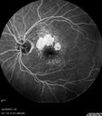
Orbital Cellulitis - Optic nerve compression with ocular ischemia557 views81 year old woman with constricted eye movement, orbital cellulitis and severe vision loss in the right eye (LP). She has whitening of the retina and the OCT shows ischemic changes as well as subretinal fluid and blood. The vision in this eye declined to NLP and the eye remained ischemic despite control of her infection.
|
|

Ocular Lymphoma - non-Hodgkins - B-Cell557 viewsMultifocal sub-RPE lesions in the left eye of a patient with history of Non-hodgkins B-cell Lymphoma last active 2 years ago
|
|

Prefoveal Opercula and early Lamellar Macular Hole557 views73 year old man with 20/32 vision and pre-foveal opercula. The OCT shows very early lamellar hole with lamellar hole epiretinal proliferation
|
|
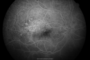
Old Branch Retinal Vein Occlusion- Severe CME - Pucker - Diabetic Early FA556 views61-year old man has diabetic retinopathy in the right eye. He also had a macular pucker and macular edema. I did a vitrectomy on September 5th. His vision was initially improving. His vision in the right eye seems to be a little more foggy.
VISUAL ACUITY: OD: 20/200
|
|

PDR with heavy PRP laser and worsening macular edema mid FA556 views51-year-old woman has had extensive laser in both eyes done years ago elsewhere. Her left eye has gradually been declining with macular edema.
VISUAL ACUITY: OD 20/40, OS 20/40
|
|
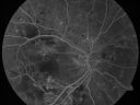
Diabetic Patient with Macular Edema and Blood Pressure 200/95556 views49-year-old decreasing vision over the last year. OD is 20/80, OS 20/80. blood pressure which was 200/95.
|
|

Proliferative Diabetic Retinopathy in patient with Moyamoya Disease556 views28-year-old womandiabetic since age nine, and she also has had multiple strokes. OD is 20/30, OS is 20/30.
|
|

Macular Fold - 40 years post retinal detachment repair - Glaucoma 556 views79-year-old woman has had a long-standing macular fold in the right eye associated with retinal detachment repair in 1970. (40 years ago) She has a macular scar in that eye. She has optic atrophy in both eyes from glaucoma. Vision OD is hand motion, OS is 20/50
|
|

Plaquenil (Hydroxycholorquine) Toxicity - Bull's Eye Maculopathy - 73 year old womand 25 Years therapy - 64 inches tall - 110 lbs556 views73-year-old woman has been on Plaquenil for twenty five years. She noticed loss of paracentral vision a few months ago. VISUAL ACUITY: OD 20/70, OS 20/30. Her color vision was 0 out 10 correct in both eyes and she also missed the controls.
|
|

Concentric Geograophic Atrophy - Age-related Macular Degeneration - Charles Bonnet Syndrome - Bulls Eye556 views88-year-old woman has age-related with vision gradually declining over the last year. She sees spots in her vision, like red spots and dark spots and she even notices during the daytime, for example when she was looking at her minister, it looked like there were little flowering pots coming out of his head.
VISUAL ACUITY: OD 20/200, OS 20/400.
|
|

Acute Annular Outer Retinopathy for 3 Months - 25 YO Female InfraRed Image556 views25-year-old female who, about three months ago, put on her sunglasses and noticed a smudge in the vision and at that point she noticed the right eye was hazy. She had a cold a week or two before the vision loss, but she didn’t have any fever. She has no focal neurological problems in her past history. VA 20/20 OU
|
|
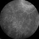
Fluorescein Angiogram - Enhanced S Cone Syndrome - Goldmann Favre - 556 views55-year-old woman while in college her vision was poor even with glasses and she sought evaluation for that. She was told after she had an electroretinogram at USF 15 years ago, that she had something with her blue cones. She does have poor night vision, but her reading vision is pretty good.
VISUAL ACUITY: OD 20/40, OS 20/40
|
|

Eccentric Disciform Scar - Breakthrough Vitreous Hemorrhage - InferoTemporal Subretinal Fibrosis - Ectopic Choroidal Neovascular Membranes556 views88-year-old man has noticed for the last few months the vision in the right eye is foggy, like there is a film in the eye or a haze, His vision is 20/16 in each eye.
Right eye - inferotemporal periphery there is a hemorrhage at the equator at about 7:00 o’clock, which is about 4-disc diameters across and adjacent to that there is scarring.
Left eye - large disciform scar inferotemporally
|
|

556 views63-year-old man diabetic for twenty two years. His central vision is fine.
VISUAL ACUITY: OD 20/25, OS 20/25
|
|

Serous Retinal Detachment - Hypertensive Retinopathy - 165/120 - Fluorescein Angiogram556 views48-year-old woman noticed when she was blinking there was a white flash in the left eye starting about a month ago. She sees it temporally. OD 20/16, OS 20/20. There is a serous retinal detachment superotemporal to the optic nerve falling short of the fovea. Her blood pressure 165/120.
|
|

Serous Retinal Detachment - Hypertensive Retinopathy -- 1 month later - less fluid (better blood pressure) (SD OCT VIDEO TAKES TIME TO LOAD)556 views48 Year old woman resolving hypertensive serous retinal detachment.
|
|

Central Serous Retinopathy 25 year old professional baseball player - Atypical - Choroidal Hypo-perfusion?556 views
|
|

Posterior Polar Annular Choroidal Dystrophy556 views68 year old woman with know macular irregularities for over 20 years. She has since been followed another 6 years and her vision remains 20/25.
|
|

Primary Ocular Lymphoma - White Mass Left Eye - 112712 556 views
|
|

Asymptomatic Juxtapapillary Choroidal Neovascular Membrane - Reticular Macular Disease (Wet AMD) 556 views
|
|

Serous Macular Detachment Both Eyes - Wet AMD vs. CSR - 62 Year Old Man - FAF556 views
|
|

Proliferative Diabetic Retinopathy - Moderate - mild NVD with NVE 556 viewsFA shows leakage from neovascularization and dark areas in periphery from non-perfusion
|
|

Retained Lens Fragments and Dislocated Intraocular Lens one week following complicated cataract surgery556 views78 year old female vision is 20/200. She developed a tear and RD during surgery for the lens fragments and has since developed PVR and had 5 surgeries. Her vision is now hand motion.
|
|

Iris Nevus556 viewsPatient came in for cataract evaluation. VA 20/200, right eye, 20/60, left eye. Slit lamp photography shows iris nevus 3-4 mm located nasally at 9-o'clock in the left eye.
|
|

Neuroretinitis - Cat Scratch - Bartonella - Sudden Vision Loss Right Eye - 29 Year Old Female556 views
|
|

Branch Retinal Vein Occlusion with NVE (Neovascularization Elsewhere)556 viewsLate Leakage on FA - NV is adjacent to non-perfusion
|
|
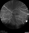
Sickle Retinopathy - Ultra Wide Field Angiogram - Heidelberg556 viewsLeakage from peripheral fronds of NV - HGB SC disease - 28 year old man with vision loss OS from acute vitreous hemorrhage
|
|

Sickle Retinopathy - Ultra Wide Field Angiogram - Heidelberg556 viewsLeakage from peripheral fronds of NV - HGB SC disease - 28 year old man with vision loss OS from acute vitreous hemorrhage
|
|

Choroidal Effusions and Serous retinal detachment with Systemic Testosterone and Cialis in 75 year old Man556 viewsOCT line scans at different visits showing the improvement of the left eye with decreasing choroidal thickness and subretinal fluid. No treatment was given.
|
|

Pre-retinal hemorrhage following Posterior Vitreous Detachment (separation - PVD)556 views68 year old woman with acute PVD OD and new preretinal hemorrhage - also there is a macular nevus
|
|

Cystoid macular edema 4 months following cataract surgery with serous retinal detachment556 views68 year old with 2 months of vision loss OS 20/100. ECCE was 4 months ago.
|
|

Orbital Cellulitis - Optic nerve compression with ocular ischemia556 views81 year old woman with constricted eye movement, orbital cellulitis and severe vision loss in the right eye (LP). She has whitening of the retina and the OCT shows ischemic changes as well as subretinal fluid and blood. The vision in this eye declined to NLP and the eye remained ischemic despite control of her infection.
|
|

Confluent Soft Drusen with Subetinal Fluid - Right Eye556 viewsAsymptomatic 85 year old man. OCT shows pocket of subfoveal fluid in the center of the macula. This could be mechanical from the drusen preventing the retina from settling down. Testing did not reveal a CNVM and subsequent OCT 6 weeks later showed no change in structure.
|
|

CRVO with Paracentral Acute Middle Maculopath - PAMM556 views56 year old woman with sudden vision loss, hemorrhages in all four quadrants. FA shows good retinal circulation. The OCT shows PAMM lesions in the affected eye. Vision did improve on second visit from 20/160 to 20/60 in about a month
|
|

Retinal Capillary Hemangioma - Endophytic and Exophytic556 views46 year old female with normal vision - her father died of pancreatic cancer
|
|

Equatorial Drusen556 views61 year old female with normal visoin
|
|

Posterior Scleritis - positive T-sign556 views62 year ols man was sick about 6 weeks ago and then 2 weeks ago the the left eye got red. He was treated for conjunctivitis and the eye did not get better. Topical steroids and oral non-steroidal anti-inflammatory medications did not help.
VA OD: Dcc20/25 NccJ5
VA OS: Dcc5'/200
The scleritis cleared within 2 weeks of starting high dose (80 mg) oral steroids. Blood tests were all negative.
|
|

Wet AMD - Subfoveal CNVM - Excellent Response to Avastin555 views76-year-old woman two to three months of vision loss OD
Vision started at 20/200 and with Avastin improved to 20/25. She has required monthly injections and as of October 2010 has been treated about 16 times. Her left eye remains good at 20/20.
The OCT from post-treatment as well as pretreatment images are in this album.
|
|
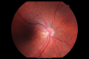
BDR OD with Edema555 views57-year-old man has diabetic retinopathy in both eyes.
Diabetic for 14 years with HgB A1C often over 10.
VISUAL ACUITY: OD 20/30, OS 20/40. PDR OS BDR OD
|
|

New BRVO Left Eye Old BRVO Right Eye - Left Eye Shows Twig Superior Vein Occlusion with Edema555 views90-year-old man decreased vision maybe one year since cataract surgery he is no longer able to read. OD 20/200, OS 20/200
|
|

Diabetic Macular Edema Left Eye - Pre Laser555 views82-year-old woman diabetic for many years, last eye exam 5 years ago with gradual vision loss. OD 20/60, OS 20/70. IOP: OD 16, OS 17.
|
|

Central Retinal Artery Occlusion less than 24 hours - 69 year old man VA light perception555 views
|
|

Ruptured Retinal Arterial Macroaneurysm - Submacular Hemorrhage - Vision 20/400 never recovered - Hypertension 205/94555 views
|
|

Branch Retinal Arterial Occlusion 20/20 Vision - Symptoms for 1 Day555 views75-year-old man was seen in the office on 4/6/2010. He noticed a sudden vision change in the right eye yesterday morning. 20/20 Vision both Eyes
|
|

Ocular Ishemic Syndrome - Bilateral Carotid Occlusions - OD 20/60 OS 20/200 Enlarged Foveal Avascular Zone and Temporal Non-perfusion555 views75-year-old man had laryngeal cancer treated in 15 year ago with radiation. He recently was told that the carotid circulation is closed off on both sides. He does have a patent vertebral artery, which is supplying him pretty well and in addition to that his cardiac output is compromised by poor mitral valves. His consideration is to what can be done about that in the future.
VISUAL ACUITY: OD 20/60, OS 20/200
|
|
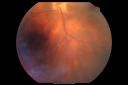
Birdshot Chorioretinitis - Retinal Vasculitis - HLA A29 Positive 555 views64-year-old woman three or four year’s fluctuations in her vision. She mostly notices them in bright light her vision glares out and she has difficulty seeing. PMHx: malaria in 1948 that was treated. She was told at one point she could possibly have had Familial Mediterranean fever.
She does intermittently get rashes around her arms. She has arthritis in her knees and ankles.
VISUAL ACUITY: OD 20/40, OS 20/50 - HLA - A29 Positive
|
|

Proliferative Sickle Retinopathy - Hemoglobin SC555 views53-year-old woman with a new spider-like floater in the right eye for about six days. She sees one big floater and another little one. She does have hemoglobin SC hemoglobinopathy. VISUAL ACUITY: Vision OD is 20/20; OS is 20/50, PH is 20/20.
|
|

Concentric Geograophic Atrophy - Age-related Macular Degeneration - Charles Bonnet Syndrome - Bulls Eye555 views88-year-old woman has age-related with vision gradually declining over the last year. She sees spots in her vision, like red spots and dark spots and she even notices during the daytime, for example when she was looking at her minister, it looked like there were little flowering pots coming out of his head.
VISUAL ACUITY: OD 20/200, OS 20/400.
|
|

Macular Pucker 20/40 Vision 555 views59-year-old woman blurry vision in the right eye for about the last one or two years. She had cataract surgery in 1981, a retinal detachment repair with scleral buckle in 1988 in the right eye, and then she had lens implants in both eyes, anterior chamber lenses, in 1994. She did start taking Latisse for eyelashes about a year and a half ago.
VISUAL ACUITY: Vision OD is 20/40, OS is 20/20
(4 months later with a short course of antiinflammatory drops and new glasses her vision improved to 20/25)
|
|

Choroidal Melanoma (Medium) Exudative Retinal Detachment 555 views87-year-old man noticed a new onset floater a few months ago. OD is 20/40, OS is 20/30
|
|
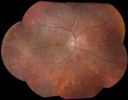
Choroidal Dystrophy555 views
|
|
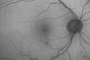
Diabetic Macular Edema - Mild555 views70 year old man with mild edema did well without treatment
|
|

Peripheral Retinal Degeneration555 viewsPatient comes in with double vision. VA was 20/20 in both eyes. Fundus exam shows Retinal Degenerative changes in both eyes. Offer to correct double vision with temporary Fresnel prism
|
|

Papilledema - Sagittal Cerebral Vein Thrombosis - Lung Transplant Cystic Fibrosis 36 Year Old Man555 viewsLine scan through optic nerve
|
|

Vitreomacular traction - recent vision loss Right Eye 555 views
|
|

Acute AION OS - Mild disc pallor OD555 viewsSudden vision loss OS x 1 week with optic nerve swelling. Evidence of optic atrophy in right eye.
|
|
| 17998 files on 180 page(s) |
 |
|
|
|
|
58 | |
|
|
|
|


