|
|

FA showing NVE, NVD, non- perfusion left eye inferonasally with neovascularization at the margin of the non-perfusion574 views
|
|
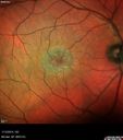
Macular Telangiectasia - Stage 3 right eye 20/40 vision574 viewsMulti-Color Image
|
|

Retinitis Pigmentosa Sine Pigmento 574 views
|
|

Polypoidal Choroidal Vasculopathy with Pulsating ICG polyp left eye574 views
|
|

Unilateral Acute Idiopathic Maculopathy (UAIM)574 views
|
|

Macular Telangiectasis (MacTel) - Blue Reflectance574 viewsGray ring is more visible before the retina is exposed to bright light. This could be from bleaching of intraretinal chromophores if they are the source of the gray ring.
|
|

HIV Retinopathy574 viewsCentral white spot is an artifact. The small vascular abnormalities are from HIV
|
|

Macular Telangiectasia and dry AMD - Simulating wet AMD574 viewsThis patient was told different things by different retina specialists. Her case is complex because she has OCT findings of Macular Telangiectasis and dry AMD. This makes it look like she has wet AMD. Her vision is stable.
|
|

Syphilitic Chorioretinitis574 viewsSevere outer retinal atrophy and RPE non-geographic atrophy from syphilis. Vision improved from 20/63 to 20/32 with therapy. Images look a lot like DUSN. Patient had several surgeries (vitrectomy for floaters, vitrectomy for macular pucker, Avastin injections and macular laser for edema) prior by other retina specialists prior to being correctly diagnosed and treated.
|
|

Retinal Capillary Hemangioma - Endophytic and Exophytic574 views46 year old female with normal vision - her father died of pancreatic cancer
|
|

Cat Scratch - Neuroretinitis OD AND retinitis OS - Bartonella henselae positive574 views27 year old female with exposure to cats and prior fever. Reports with recent vision loss right eye. She was treated with doxycycline and within 2 weeks the right eye looks better. Then she skipped all follow-up visits. The right eye presented 20/200 and the left 20/16
|
|

Type 1 Retinal Arteriovenous Malformation574 views42 year old female VA 20/32 OD, 20/20 OS MRI was negative.
|
|
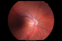
BDR OD with Edema573 views57-year-old man has diabetic retinopathy in both eyes.
Diabetic for 14 years with HgB A1C often over 10.
VISUAL ACUITY: OD 20/30, OS 20/40. PDR OS BDR OD
|
|

Diabetic Retinopathy - Non-Perfusion - Possible Central Retinal Vein Occlusion573 views68-year-old woman OD 20/70, OS 20/200 CYSTOID MACULAR EDEMA, POSSIBLE EITHER OCULAR ISCHEMIC SYNDROME IN THE RIGHT EYE OR OCCULT CENTRAL RETINAL VEIN OCCLUSION IN THE RIGHT EYE,S EVERE NON-PerfUSION WITH DIABETIC RETINOPATHY IN THE LEFT EYE.
|
|

BDR and Diabetic Macular Edema with Fresh Laser Right Eye573 views60-year old man OD 20/60, OS 20/50 worsening vision both eyes
|
|

Toxoplasmosis - Inner Retinal - Recurrent - Treated with Bactrim DS 573 views43-year-old man has macular toxoplasmosis. He has responded nicely to Bactrim OS: 20/60
|
|

Hypertensive Retinopathy with Choroidal Ischemia Right Eye and Vision 8/200 - Blood Pressure 240/120 mmHg IgA Nephropathy573 views39-year-old man two days ago he noticed poor vision in his right eye OD: 8/200; OS: 20/20. BP is 240/120
|
|

Proliferative Diabetic Retinopathy in patient with Moyamoya Disease573 views28-year-old womandiabetic since age nine, and she also has had multiple strokes. OD is 20/30, OS is 20/30.
|
|

Central Retinal Artery Occlusion less than 24 hours - 69 year old man VA light perception573 views
|
|

Central Retinal Artery Occlusion less than 24 hours - 69 year old man VA light perception573 views
|
|

Ruptured Retinal Arterial Macroaneurysm - Submacular Hemorrhage - Vision 20/400 never recovered - Hypertension 205/94573 views
|
|

Hypertensive Choroidopathy - Pheochromocytoma - Chronic Very High Blood Pressure 49 YO Male573 views49-year-old man chronic very high blood pressure sometimes the blood pressure will go as high as 260/160, sometimes it is 200/120 and sometimes it is 160/80. He has a pheochromocytoma on the adrenal gland and he has had vision loss in the right eye starting between three and four years ago.
VISUAL ACUITY: OD 20/60, OS 20/25.
|
|

573 views
|
|

Geographic Atrophy 20/160 Vision573 views84-year-old woman has age-related macular degeneration in both eyes with foveal atrophy. She also has a central retinal vein occlusion
in the left eye for which she had intravitreal Kenalog and finally chorioretinal anastomosis laser a year
ago. VISUAL ACUITY: Vision OD is 20/160, OS is 7/200.
|
|

Central Serous Retinopathy - Steroid Induced - Serous Retinal Detachment573 views38-year-old man has noticed a spot in the right eye. The vision was a little bit off for a few weeks and then just recently he noticed that it was the right eye. He did take high dose oral steroids for about two weeks in early March and he was using an intermittent steroid inhaler, but he has not used anything recently. He is under a lot of stress.
VISUAL ACUITY: OD 20/100, OS 20/50.
|
|

VPRT573 views
|
|
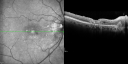
Juxtafoveal Telangiectasis (MacTel) and Asymptomatic Extrafoveal Juxtapapillary Choroidal Neovascular Membrane573 views84-year-old woman has juxtafoveal retinal telangiectasis in both eyes. The right eye has an asymptomatic extra foveal juxtapapillary choroidal neovascular membrane. OD 20/400, OS 20/63.
|
|

Equatorial Drusen573 views76-year-old man with equatorial drusen
VISUAL ACUITY: Vision OD is 20/40, OS is 20/25.
|
|

Giant Retinal Tear Initial Presentation573 views61 yo phakic male with giant retinal tear OD
|
|
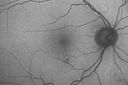
Diabetic Macular Edema - Mild573 views70 year old man with mild edema did well without treatment
|
|

Central Serous Retinopathy 25 year old professional baseball player - Atypical - Choroidal Hypo-perfusion?573 views
|
|

Asymptomatic Juxtapapillary Choroidal Neovascular Membrane - Reticular Macular Disease (Wet AMD) 573 views
|
|

Reticular Macular Disease (Pseudo-drusen) Both Eyes - Occult CNVM and Macular Edema Right Eye 573 viewsIndocyanine Green Angiogram
|
|
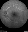
Macular Pucker 20/40 vision - minimal symptoms for 1 year Right eye573 viewsFA shows leakage from traction from pucker in the right eye - cystoid macular edema
|
|

Acute non arteritic anterior ischemic optic neuropathy left eye - SD Optical Coherence Tomography Line Scan573 views66 y.o. woman spot in central vision in left eye started about 1 week ago since then it is not changing. VA OD: sc20/25 OS: sc20/200
|
|

Retained Lens Fragments and Dislocated Intraocular Lens one week following complicated cataract surgery573 views78 year old female vision is 20/200. She developed a tear and RD during surgery for the lens fragments and has since developed PVR and had 5 surgeries. Her vision is now hand motion.
|
|

Branch Retinal Vein Occlusion with worse macular edema from hypertension - FAF Photo from 2014 - vision 20/40573 views
|
|

Acute Posterior Multifocal Placoid Pigment Epitheliopathy - 4 years after onset573 views4 years after presentation - vision 20/80 OD 20/16 OS and there is subretinal fibrosis
|
|

Syphillitic Optic Neuropathy and Maculopathy573 views46 year old woman with 2 weeks of vision loss OD. VA 20/200 OD; 20/20 OS. there are posterior vitreous cells on OCT in both eyes. ICG, FAF, and FA show an abnormal macula OD. VA improved to 20/30 within a month with treatment with PCN.
|
|

Macular Edema and dry AMD - excellent example of why FA and ICG are necessary in management of wet AMD573 viewsThis 72 year old woman has an OCT that looks like wet AMD. Only her FA and ICG show that she has inflammatory macular edema. She had no clinical signs or symptoms of uveitis. Nevertheless, the macular edema responded to subtenon's kenalog.
|
|

Orbital Cellulitis - Optic nerve compression with ocular ischemia573 views81 year old woman with constricted eye movement, orbital cellulitis and severe vision loss in the right eye (LP). She has whitening of the retina and the OCT shows ischemic changes as well as subretinal fluid and blood. The vision in this eye declined to NLP and the eye remained ischemic despite control of her infection.
|
|

Birdshot Chorioretinitis - Fresh Presentation573 viewsUltawidefield ICG shows best the multifocal lesions. 61 year old woman with floaters for less than 6 months. HLA-A29 Positive
|
|

Coats' Disease - 14 year old - Asymptomatic573 viewsPresented with macular exudates. Temporal macroaneurysms responded to laser. Ultra-wide field FA shows nonperfusion
|
|

Syphilitic Chorioretinitis573 viewsSevere outer retinal atrophy and RPE non-geographic atrophy from syphilis. Vision improved from 20/63 to 20/32 with therapy. Images look a lot like DUSN. Patient had several surgeries (vitrectomy for floaters, vitrectomy for macular pucker, Avastin injections and macular laser for edema) prior by other retina specialists prior to being correctly diagnosed and treated.
|
|

Syphilitic Chorioretinitis573 viewsSevere outer retinal atrophy and RPE non-geographic atrophy from syphilis. Vision improved from 20/63 to 20/32 with therapy. Images look a lot like DUSN. Patient had several surgeries (vitrectomy for floaters, vitrectomy for macular pucker, Avastin injections and macular laser for edema) prior by other retina specialists prior to being correctly diagnosed and treated.
|
|

Optic Nerve Pit Retinal Detachment - SD OCT of Nerve EDI shows Pit573 views9 Year old with morning glory disc who developed nerve pit RD. Raster scans of the optic nerve on EDI setting show the communication between the subretinal space and the fluid around the cerebrospinal fluid surrounding the optic nerve sheath.
|
|
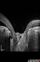
Optic Nerve Pit Retinal Detachment - SD OCT of Nerve EDI shows Pit573 views9 Year old with morning glory disc who developed nerve pit RD. Raster scans of the optic nerve on EDI setting show the communication between the subretinal space and the fluid around the cerebrospinal fluid surrounding the optic nerve sheath.
|
|

Proliferative Diabetic Retinopathy - NVD regressed with PRP laser573 views76 year old diabetic man - Presented one year ago with NVD in the left eye. This regressed with laser but then a year later worsened. Additional PRP was done and the NVD regressed again.
|
|

Foveal hypoplasia - aplasia573 viewsSD OCT shows persistent plexiform layers in the center of the macula
EXTENDED HPI: He is having some trouble seeing his gadgets (like his watch). He was diagnosed with morning glory syndrome at about 3 yeras old. This last visit you saw fluid under the retina and asked him to come here. The vision is a little worse.
VA OD: Dcc20/40+2 NccJ2-2
VA OS: Dcc20/25-2 NccJ1-2
|
|

Subtle Branch Retinal Artery Occlusion573 views68 year old man with shadow in the left eye yesterday. IR and OCT show subtle retinal swelling. FA is normal
|
|

Branch retinal vein occlusion with macular edema573 views71 year old man Starting about 2 weeks ago the left eye was blurred. The right eye is seeing well.
VA OD: sc20/25+2 NccJ1+
VA OS: sc20/63+2 PH20/50-2 Ncc20/400
IOP: TP: OD:19 OS:16
|
|
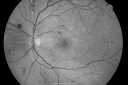
BDR and Diabetic Macular Edema with Fresh Laser Right Eye572 views60-year old man OD 20/60, OS 20/50 worsening vision both eyes
|
|

Multiple Evanescent White Dot Syndrome - Acute Idiopathic Blind Spot Enlargement572 views33-year-old woman one week ago she awoke and saw a red spots flashing out of her right eye. She notices them to the right side, which is the temporal visual field out of the right eye, then they changed to bursts of white lights without colors and then she notices straight ahead a couple spots. She has headaches in the back of her head. OD 20/20, OS 20/20
|
|

Choriodal Neovascular Membrane - Subfoveal - Pseudo-Retinitis Pigmentosa - 50 years post Retinal Detachment Repair572 views86-year-old with a history of decreased vision in the right eye. The patient reports she has lost vision in the right eye over the past two months or so. She reports the vision was always compromised in that eye since retinal detachment surgery 50 years ago with a scleral buckler. Vision dropped from 20/50 to 20/200 right eye in 2 months.
|
|

Macular Hemorrahge and Probable CNVM Right eye for 2 weeks VA 20/400 - 20 Year old572 views20-year-old man noticed vision loss in the right eye about two weeks ago and if anything it seems to be getting worse. He is a high myope. VISUAL ACUITY: OD 20/400
|
|

Macular Pucker - 3 Weeks Vision Loss - 20/50 Vision - Preoperative Images572 viewsPREOP:
59-year-old man new onset floaters about a year ago. Subsequent to that his vision was not quite right in the right eye and then he noticed, starting about three weeks ago, substantial change in the vision. VISUAL ACUITY: Vision OD is 20/50, OS is 20/16
|
|

Macular Pucker - 3 Weeks Vision Loss - 20/50 Vision - Preoperative Images572 viewsPREOP:
59-year-old man new onset floaters about a year ago. Subsequent to that his vision was not quite right in the right eye and then he noticed, starting about three weeks ago, substantial change in the vision. VISUAL ACUITY: Vision OD is 20/50, OS is 20/16
|
|

Non-arteritic Anterior Ischemic Optic Neuropathy following Intravitreal Injection 572 views81-year-old woman has had injections in both eyes for wet age-related macular degeneration. 2 weeks ago the left eye was treated. Less than a week ago she noticed a change in vision in the left eye. She had a dull left frontal headache and noticed her vision was hazy as if there was a veil over the vision. OD is 20/25, OS is 20/25. IOP: OD 18, OS 26. The left eye has a 3+ APD
|
|

Macular Branch Retinal Vein Occlusion and Adult Vitelliform Dystrophy572 views72-year-old woman 2 months post ECCE with poor vision since surgery especially left eye.
VISUAL ACUITY: OD 20/50, OS 20/200
|
|

Gonioscopy, Blood in the Anterior Chamber from Hyphema572 viewsPatient comes in with blunt trauma to the right eye due to a BB gun incident. Patient was present with a hyphema at 8-o'clock about 1mm thick. Gonioscopy photos were then taken to show blood from the hyphema entered into the anterior chamber. Patient had no angle recession in the right eye.
|
|

PDR572 views
|
|

Vitreomacular traction - recent vision loss left eye - FA572 views
|
|

Free Floating Dislocated Lens in Vitreous572 viewsPatient comes in aphakic with dislocated lens floating to the back of the eye when laying down. Lens is laying up against the endothelium of the cornea when patient is right side up..
|
|

Acute AION OS - Mild disc pallor OD572 viewsSudden vision loss OS x 1 week with optic nerve swelling. Evidence of optic atrophy in right eye.
|
|

Cystoid macular edema 4 months following cataract surgery with serous retinal detachment572 views68 year old with 2 months of vision loss OS 20/100. ECCE was 4 months ago.
|
|

Cystoid Macular Edema 10 years after Cataract Surgery572 viewsNo overt uveitis but the patient had aching in the eyes and responded to topical anti-inflammatory drops with recurrence of the CME on discontinuation of topical therapy
|
|

CRVO with Paracentral Acute Middle Maculopath - PAMM572 views56 year old woman with sudden vision loss, hemorrhages in all four quadrants. FA shows good retinal circulation. The OCT shows PAMM lesions in the affected eye. Vision did improve on second visit from 20/160 to 20/60 in about a month
|
|
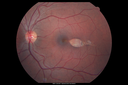
Torpedo Maculopathy572 viewsLikely a variant of CHRPE
|
|

Myelinated Nerve Fiber Layer - Optic Nerve572 viewsRight eye subtle and inferior - left eye obvious
|
|

Chronic CSC Both Eyes - Recent vision loss Right Eye Treated with PDT572 views50 year old man The right eye has had vision loss for a few weeks. He had previous episodes seen by me in 2012 OS and 2008 OD. This was 2018. VA OS has been poor for several years.
VA OD: Dcc20/100
VA OS: Dcc20/200-3
The patient had PDT to leaky area superotemporal to the fovea 1/2 dose and within 3 months was back up to 20/25. The last two OCT scans show fluid absorbing
|
|

Posterior Scleritis - positive T-sign572 views62 year ols man was sick about 6 weeks ago and then 2 weeks ago the the left eye got red. He was treated for conjunctivitis and the eye did not get better. Topical steroids and oral non-steroidal anti-inflammatory medications did not help.
VA OD: Dcc20/25 NccJ5
VA OS: Dcc5'/200
The scleritis cleared within 2 weeks of starting high dose (80 mg) oral steroids. Blood tests were all negative.
|
|
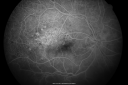
Old Branch Retinal Vein Occlusion- Severe CME - Pucker - Diabetic Early FA571 views61-year old man has diabetic retinopathy in the right eye. He also had a macular pucker and macular edema. I did a vitrectomy on September 5th. His vision was initially improving. His vision in the right eye seems to be a little more foggy.
VISUAL ACUITY: OD: 20/200
|
|
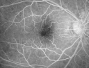
Cystoid Macular Edema - Xalatan - ECCE was done 20 years previously571 views71 Year old woman who noticed about a month ago the central vision in the right eye fogged up. OD 20/200 POSSIBLE XALATAN INDUCED CYSTOID MACULAR EDEMA
|
|

Bilateral Retinal Arteriol Occlusions - Possible Susac Syndrome 571 views80-year-old woman one month ago had vision loss and a vascular occlusion in the right eye. Vision loss occurred in the left eye about 9 years ago with cotton wool spots. Patients has a 30 year history of tinnitis.
|
|

Patterned Pigment Dystrophy of the Fovea 79 YO Woman VA 20/20 Right 20/30 Left571 views
|
|

Concentric Geograophic Atrophy - Age-related Macular Degeneration - Charles Bonnet Syndrome - Bulls Eye571 views88-year-old woman has age-related with vision gradually declining over the last year. She sees spots in her vision, like red spots and dark spots and she even notices during the daytime, for example when she was looking at her minister, it looked like there were little flowering pots coming out of his head.
VISUAL ACUITY: OD 20/200, OS 20/400.
|
|

Cystoid Macular Edema - Xalatan - ECCE was done 20 years previously - 1 year later 20/40 Vision571 viewsXalatan stopped and a brief course of topical anti-inflammatory drops was prescribed. One year later vision is back to normal
|
|

Macular Pucker 20/40 Vision 571 views59-year-old woman blurry vision in the right eye for about the last one or two years. She had cataract surgery in 1981, a retinal detachment repair with scleral buckle in 1988 in the right eye, and then she had lens implants in both eyes, anterior chamber lenses, in 1994. She did start taking Latisse for eyelashes about a year and a half ago.
VISUAL ACUITY: Vision OD is 20/40, OS is 20/20
(4 months later with a short course of antiinflammatory drops and new glasses her vision improved to 20/25)
|
|

Choroidal Melanoma - Exudative Retinal Detachment 82 Year Old Man 571 views82-year-old man who had 3 falls 2 weeks ago. After the falls he started checking his vision and noticed there was a veil over the left eye, which he had seen for about two weeks in the superior visual field. OD 20/25, OS 20/40
(patient was medically never well enough for brachytherapy and died 6 months later from heart disease)
|
|

Retinitis Pigmentosa Newly Diagnosed 55 Year Old571 views55-year-old noticed his vision declining recently, over the last few weeks, particularly in the right eye. He said previous to that the vision was not normal but was pretty good. OD is 20/100, OS is 20/50
OCT SCAN: There is macular thickening in both eyes with intraretinal cysts.
|
|
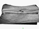
Group 2a Juxtafoveal Telangiectasis - MacTel - Stage 5 left eye - Macular Hole Right eye571 views76-year-old woman has juxtafoveal retinal telangiectasis. She had Stage five neovascularization in the left eye I treated with injections between September of 2007 and October of 2008 with Avastin (3 years ago). OCT scan today shows macular hole right eye. VISUAL ACUITY: OD 20/400, OS 20/40.
|
|

Cuticular Drusen (Basal Laminar Drusen) and Bilateral Chronic Subfoveal Fluid (Serous Retinal Detachment) unresponsive to monthly Avastin Infrared OD571 views66-year-old man with bilateral subfoveal fluid unresponsive to one year of monthly avastin therapy.
OD 20/160, Pinhole 20/80. OS 20/63, Pinhole 20/50. OCT scan shows subretinal fluid with normal choroidal thickness. Fluorescein angiogram shows cuticular/basal laminar drusen. ICG shows no foci of choriocapillaris leakage.
|
|

Hyperpigmentation of the Retinal Pigment Epithelium Right Eye (Treated wet AMD OS)571 views85-year-old man has wet age-related macular degeneration in the left eye treated for one year most recently one year ago. OD is 20/30, OS is 20/60
|
|
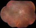
Choroidal Dystrophy571 views
|
|
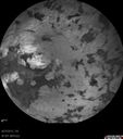
Retinitis Pigmentosa - Autosomal Dominant - Vision Loss at Young Age - Infrared571 views 54-year-old woman has autosomal dominant retinitis pigmentosa. Her mother and grandmother both had it and she has one son who has it and one daughter who doesn’t. She also has a sister with it. She lost vision since she was younger. She had cataract surgery done in January and February of this year. She has been seeing unusual flashing lights throughout the periphery and central vision for about the last year or two.
VISUAL ACUITY: OD 20/160, OS 20/160
|
|

Pigmented Peripheral Retinal Degeneration571 views42-year old male comes in for routine eye exam and to follow up on peripheral retinal degeneration in both eyes. VA is 20/20, right eye and 20/25, left eye. Patient is asymptomatic with no visual complaints.
|
|

BDR571 views
|
|

Serous Macular Detachment Both Eyes - Wet AMD vs. CSR - 62 Year Old Man - Color Photo571 views
|
|

571 views
|
|

Choroideremia - Female Carrier - Patchy Atrophy of Choroid and RPE - Lyonization571 views
|
|

Posterior Uveal Effusion571 views
|
|

IPCV - idiopathic polypoidal choroidal vasculopathy - juxtapapillary CNVM with hemorrhage both eyes (also choroidal nevus right eye) - Subretinal hemorrhage both eyes571 views86 year old woman with shadow in vision OD and new hemmorhage OU.
|
|

Acute Posterior Multifocal Placoid Pigment Epitheliopathy - 4 years after onset571 views4 years after presentation - vision 20/80 OD 20/16 OS and there is subretinal fibrosis
|
|
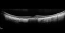
Central Areolar Choroidal Dystrophy571 viewsVision loss from early 60's. This 78 year old woman has choroidal atrophy centrally.
|
|

Macular Laser Scars Left Eye - 5/200 Vision571 views83 year old woman who had extensive macular laser in the left eye about 15 years ago for DME. Vision is now 20/50 right eye and 3/200 left eye
|
|

Subretinal Hemorrhage - Commotio Retinae - Macula - hit with baseball571 views15 year old with VA 20/60 3 days after being hit with baseball in the right eye
|
|

Pre retinal hemorrhage associated with partial PVD571 views56 year old female in for checkup- no symptoms and normal vision - there is a hemorrhage by the nerve that could be vascular, tractional or from her mild glaucoma (which is controlled). The macular hemorrhage is preretinal.
|
|

Macular Telangiectasis - Very Mild571 views52 year old female with normal vision and suspicious OCT referred. Multimodal imaging shows very mild mactel (loss of macular pigment, ring on multicolor image and very mild OCT findings)
|
|

Moderate PDR571 views43 year old man 20/32 vision in both eyes. Being started on Anti-VEGF injections which will be followed by PRP.
|
|

Vitreomacular traction - spontaneous release571 views80 year old man with transient vision loss from 20/20 - 20/30 from vitreomacular traction. No surgery or procedure was done.
|
|
| 18117 files on 182 page(s) |
 |
|
|
|
|
59 | |
|
|
|
|


