|
|

Retinal Arterial Macroaneurysm - Normal Vision (2 Years before rupture)1086 views78-year-old woman has a retinal arterial macroaneurysm in the right eye. VA 20/25 (pre-rupture photos)
|
|
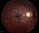
Acute Multifocal Placoid Pigment Epitheliopathy (AMPPE) related scarring1083 views
|
|

Macular Pucker1083 viewsPatient comes in for followup on macular pucker, right eye. VA is 20/50 in the right eye...Fundus photo shows prominent pucker centrally pulling temporally in the right eye.
|
|

Patterned Pigment Dystrophy of the Fovea 2 year later VA still good1082 views
|
|

Silicone Oil in Anterior Chamber and Band Keratopathy 8 Years Post Complex Retinal Detachment Repair1082 views81-year-old man had complex retinal detachment repairs in both eyes with silicone oil 8 years ago. The left eye has an oil fill. He had a retinotomy very near to the macula and he has had silicone oil in the eye for a few years. His eye is comfortable and the vision is stable. OD is 20/200, OS is light perception
|
|

Ectopia Lentis - Marfan's Syndrome - Intermittent Pupillary Block Glaucoma Right Eye1081 views41-year-old woman decreased vision right eye and intermittent pupillary block glaucoma for 6 months from a dislocated natural lens in the eye, probably associated with Marfan syndrome.Vision OD is 20/160, PH 20/40; OS is 20/30, PH 20/25
|
|

Papilledema - Spectral Domain OCT used for Diagnosis - 14 year old Child - FAF1081 views14-year-old was with optic nerve swelling asymptomatic - picked up during routine eye exam. OD 20/20, OS 20/32
SD-OCT is used to differentiate optic disc edema from optic nerve head drusen (which are not yet calcified in children).
|
|

Acute Central Serous Retinopathy - Classic Smokestack on Fluorescein Angiogram 1080 views34-year-old man noticed about five days ago decreased vision in the right eye. He was having difficulty playing video games and seeing with that eye. He thought maybe his refractive error was shifting.
VISUAL ACUITY: Vision OD is 20/40, OS is 20/16
|
|

Phacoantigenic Reaction1079 viewsPicture shows a Left eye enucleation with H&E staining.
Anterior capsule of lens is broken with evidence of granulomatous inflammation.
|
|

Mild Proliferative Diabetic Retinopathy1079 viewsRight eye has NVE and left eye has NVD and NVE. VA 20/25 OU.
|
|

Fundus Flavimaculatus - Stargardt Disease - 20/50 OD 20/200 OS 61 Year old Infrared Image OD1078 views61-year-old decreasing vision for about the last five years. OD 20/50, OS 20/200.
Pisciform Lesions and Macular Atrophy
|
|

Purtchers Retinopathy with Bilateral Central Retinal Artery Occlusions 1077 views50-year-old man traumatic chest compression and leg crushing injury. OD 1/200, OS 3/200
|
|

Acute Central Serous Retinopathy - Classic Smokestack on Fluorescein Angiogram 1077 views34-year-old man noticed about five days ago decreased vision in the right eye. He was having difficulty playing video games and seeing with that eye. He thought maybe his refractive error was shifting.
VISUAL ACUITY: Vision OD is 20/40, OS is 20/16
|
|

Central Retinal Vein Occlusion1076 viewsPatient comes in with mild blurred vision in the right eye. Fundus exam shows CRVO with scattered retinal hemorrhages in the right eye.
|
|

Plaquenil Toxicity - Bulls Eye Maculopathy1075 views70-year-old woman with systemic Lupus erythematosus and clotting problems. She was on the Plaquenil for about eight years and then off the Plaquenil for the last eight years because she developed macular toxicity. Although her vision was hazy, it was stable. Recent deceased vision left eye: OD 20/60, OS 20/100. IOP: OD 18, OS 19.
|
|

Patterned Pigment Dystrophy of the Fovea 79 YO Woman VA 20/20 Right 20/30 Left1075 views
|
|

Central Serous Retinopathy Acute - Fundus Autofluorescence1074 views42-year-old man was seen in the office on October 5, 2011. He had noticed starting in August after a course of antibiotic and steroids, that he developed new spots in his vision in the right eye. He may have had an episode like this sometime in the past. He did take steroids a few years ago and his vision did change at that time, but then returned.
VISUAL ACUITY: OD 20/32, OS 20/32
|
|
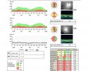
Optic Nerve Head Drusen Nerve Fiber Layer Scan1073 views57-year-old woman has optic nerve drusen in both eyes.
VISUAL ACUITY: OU 20/20.
|
|

Central Serous Retinopathy Acute 1073 views42-year-old man was seen in the office on October 5, 2011. He had noticed starting in August after a course of antibiotic and steroids, that he developed new spots in his vision in the right eye. He may have had an episode like this sometime in the past. He did take steroids a few years ago and his vision did change at that time, but then returned.
VISUAL ACUITY: OD 20/32, OS 20/32
|
|

CHRPE lesion in the left eye - Irregular pigmentation1072 views63 year old female with normal vision and CHRPE lesion in the right eye.
|
|

Optic Nerve Drusen in 7 Year Old Child - Miminal Calcification1072 views7-year-old child difficulty seeing the blackboard in school. She was checked for glasses and it was noted at that time that she had unusual looking optic nerves. Vision OU is 20/50.
|
|

Multiple Evanescent White Dot Syndrome - Atypical Yellow Foveal Spot Right Eye1071 views12-year-old had decreased vision in the left eye from reported amblyopia since around the year 2000. His right eye was doing fine until two days ago, when he had sudden severe vision loss. He sees a blurred spot in the middle of the vision in the right eye wherever he looks.
OD 20/60 with eccentric movement, OS 20/200. Pinhole 20/70. IOP: OD 14, OS 10.
|
|

Gonioscopy; Scattered Peripheral Anterior Synechiae1071 viewsPatient comes in for evaluation for glaucoma. Patient also has a history of Uveitis. Last flare up was back in 1990. Patient's VA was 20/30, Right eye and 20/40-1, Left eye. Slit Lamp Gonioscopy reveals iris bow with scattered PAS around the angles of the anterior chamber. You can also see pigmentation in the trabecular meshwork. Patient will follow up in 3-months.
|
|

Proliferative Diabetic Retinopathy - Moderate - mild NVD with NVE 1071 viewsVenous beading, vascular loops and NVE are visible on photos
|
|

Gonioscopy- Pigmentary Glaucoma Suspect1070 viewsPatient with no family history of glaucoma, comes in with elevated IOP. During Gonioscopy exam. brown pigment overlying the trabecular meshwork. Also, trans-illumination defects on the iris.
|
|

Ahmed Tube Placement1069 viewsPatient with severe glaucoma is seen post op for ahmed tube placement. VA is 20/70 and IOP is 8 in the left eye. Patient is stable.
|
|

Iris Nevus1069 viewsRoutine eye exam. Iris nevus located around 4-o'clock off the pupil.
|
|

Dislocated Intraocular Lens1068 viewsDislocated Intraocular Lens
|
|

Commotio Retinae - One Day following Basketball to Eye1068 views43-year-old man was playing basketball last night. He was hit straight in the left eye with the ball. Initially he had darkness in his inferior vision and now he has darkness inferonasally in the left eye.
Vision OD is 20/16, vision OS is 20/32
|
|

Radioactive Plaque Placement for Choroidal Melanoma Eye is Anesthetized Prior to Surgery1067 views
|
|

Malignant Hypertension - Cotton Wool Spots - Elschnig Spots - Optic Nerve Edema 1066 views
|
|

Calcified Drusen - Dry Age-related Macular Degeneration - Geographic Atrophy1065 views87 Year old woman with dry age-related macular degeneration in both eyes.With both eyes open she is fine, but when she closes the left eye, things are wavier.
VISUAL ACUITY: Vision OD is 20/100, OS is 20/40.
|
|

Progressive Outer Retinal Necrosis 77 Year Old Woman with CLL (Acute Retinal Necrosis) (PORN - ARN)1064 views77-year-old woman with CLL who had shingles on the left side of her face about 6 weeks ago then she developed a dendrite in the cornea which was treating about four weeks ago. She noticed severe vision loss in the left eye just a few days ago and you saw retinitis and she comes in because of that. Vision OD is 20/25, OS is hand motion
|
|

Giant Papillary Conjunctivitis, Left Upper Eyelid1064 viewsContact lens wearer, comes in for exam. Has rough feeling underneath both eyelids. Patient thought it was through SCL wear. Patient VA was 20/20. right eye, 20/30, left eye. Underneath the left upper eyelid, you can see papillary inflammation and redness.
|
|

Gonioscopy, Pigment Dispersion Syndrome1064 viewsPatient comes in with elevated pressures. Goinoscopy shows heavy pigment dusted off the iris into the trabecular meshwork. Laser procedure was done to break up the pigment for more aqueous flow.
|
|

Mild Clinically Significant Diabetic Macular Edema1063 views58-year-old woman diabetic for 20 years who has diabetic macular edema in both eyes. She has had waxing and waning edema in the past and I thought it might improve on its own but it did not. She notices her vision is still a little hazy.
VISUAL ACUITY: Vision OD is 20/30, OS is 20/25.
|
|
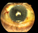
Ozurdex Implant in AC1063 views79yr old male presents Blurry vision with slight discomfort x 1 week in OD . Pt is S/P Ozurdex Implant x 2mos which is now surfaced in the Anterior Chamber.
|
|

Vitreo-Traction due to Diabetic Retinopathy1063 viewsPatient comes in for follow up on her diabetes. VA is 20/30 in the right eye. Fundus photos shows thick traction nasally to the optic nerve forming a ring around the macula. Will follow up in 6-months.
|
|

Flower Cataract1061 viewsPatient with ahmed tube and flower cataract
|
|

Macular Degeneration with Hemorrhage1060 viewsPatient comes in for follow up exam for wet AMD. Fundus photography reveals retinal hemorrhage temporally to the macula. Patient was not treated. Wait to see if the blood will absorb on its own.
|
|

Central Areolar Choroidal Dystrophy1059 viewsVision loss from early 60's. This 78 year old woman has choroidal atrophy centrally.
|
|

Three years after Injury - Optic Atrophy - Vision is 1/200 in each eye1058 views
|
|

Conjunctival Melanoma1058 viewsRight conjunctival melanoma extending into anterior orbit, right eye. Temporally and nasally with pigmented masses/nodules. VA was 20/30 without correction in the right eye. Follow up to proceed with proton beam therapy.
|
|

Hyperpigmentation of Retinal Pigment Epithlium in Dry AMD (hyperplasia)1057 views87-year-old woman has wet age-related macular degeneration in the left eye. She had treatment up north but there has been a gap in therapy and the vision in the left eye is poor. Vision OD is 20/25, OS is 20/400
|
|

Fundus Flavimaculatus - Stargardt Disease - 20/50 OD 20/200 OS 61 Year old Color Photo1057 views61-year-old decreasing vision for about the last five years. OD 20/50, OS 20/200.
Pisciform Lesions and Macular Atrophy
|
|
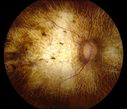
Chorodial Sclerosis1057 views74 year old female diagnosed with Chorodial Sclerosis OU and ARMD OU. Va 20/400 OD 2'200 OS
|
|

Diabetic Retinopathy with CME1054 viewsSecond opinion for diabetic retinopathy. VA is 20/30, right eye, 20/50, left eye. Patient has mild swelling in the macula of the right eye. No evidence of CME but does have a moderate cataract in the left eye.
|
|

Acute Zonal Occult Outer Retinopathy1053 views45-year-old man noticed a few years ago peripheral vision blurriness in the left eye and also some central vision loss. Previous to that, as far as he knows, the two eyes were okay.
VISUAL ACUITY: OD: 20/20; OS: 20/40.
|
|

Optic Pit OCT1053 viewsOCT Scan of an Optic Pit in OS of a 40 yr old male
|
|

Hemicentral Retinal Vein Occlusion Vision 20/60 1052 views84-year-old man has a hemicentral retinal vein occlusion in the right eye. His vision has declined since then.
VISUAL ACUITY: Vision OD is 20/60.
|
|

Cilia and Cement Intraocular Foreign Body1052 viewsConstruction worker jack-hammering 3 months prior to pictures. Asymptomatic. 20/20 OU. Corneal wound seen superiorly (not in pictures)
|
|

Sturge-Weber Encephalotigeminal Angiomatosis - Facial Hemangioma and Asymptomatic Ipsilateral Diffuse Choroidal Hemangioma1051 views61-year-old man with Sturge-Weber syndrome with a hemangioma on the left side of his face.
VISUAL ACUITY: Vision OD is 20/50, PH 20/30; OS 20/80, PH 20/30. IOP: OD 16, OS 19.
|
|

New Subfoveal Classic CNVM OS - Wet AMD - Macular Scar Congenital Toxoplasmosis Right Eye 1050 views77-year-old woman as had poor vision in the right eye since birth from congenital toxoplasmosis of her macula in the right eye. Two to three weeks ago when she noticed vision loss in the left eye. VISUAL ACUITY: OD 2/200, OS 20/200.
|
|

Equatorial Drusen Fundus Photo Left Eye1049 views
|
|

Retinocytoma - Retinoma - Regressed Retinoblastoma 43 year old male incidental finding 1049 views
|
|

Optic Nerve Drusen - - Nerve Fiber Layer Scan1049 views72-year-old woman has had optic nerve drusen for sometime and has visual field abnormalities. OD 20/32, OS 20/32.
|
|

wyburn-mason syndrome fundus photos1048 views28 year old female, visual acuity 20/200
|
|

PDR with heavy PRP laser and worsening macular edema 1046 views51-year-old woman has had extensive laser in both eyes done years ago elsewhere. Her left eye has gradually been declining with macular edema.
VISUAL ACUITY: OD 20/40, OS 20/40
|
|

Pigmented Peripheral Retinal Degeneration1045 views42-year old male comes in for routine eye exam and to follow up on peripheral retinal degeneration in both eyes. VA is 20/20, right eye and 20/25, left eye. Patient is asymptomatic with no visual complaints.
|
|

Optic Nerve Hypoplasia1042 views28-year-old man with diabetes and anomalous vessels near nerve. OD is 20/25, OS is 20/20. IOP: OD 20, OS 22.
OD: Vertical C/D ratio is 0.0. The nerve is tilted and small and there is an anomalous branching pattern of the major retinal vessels. There is a tuft of irregular telangiectatic vessels on the nasal edge of the optic nerve. There is also a buried optic nerve druse at the optic margin at 5:00 o’clock. There are rare retinal hemorrhages in the macula.
OS: Vertical C/D ratio is 0.0. Again, the nerve looks small. There is an anomalous branching pattern of the retinal vessels and rare retinal hemorrhages.
|
|
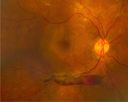
PDR with subhyloid hem1042 viewsPDR with subhyloid hem
|
|

Accordioning Crystalline Lens with loss of Posterior Capsule1042 views71-year old male complains of blurred vision in the left eye. VA 20/40, right eye and 20/400, left eye without correction. Slit lamp exam shows Crystalline lens, both eyes. Right eye IOL is aligned and centered. Left eye shows an accordion of the crystalline lens. Retro Illumination shows the IOL bent inward in the left eye. There was a 6-diopter difference of astigmatism between the right and left eye. Patient will have surgery to correct the issue.
|
|
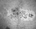
Bilateral Diffuse Uveal Melanocytic Proliferation - BDUMP - Paraneoplastic Syndrome1041 views80-year-old man vision loss for one year. He died about one year after these photos from Metastatic Poorly Differentiated Large Cell Carcinoma of unknown primary. He was a smoker.
|
|

Occult Maculopathy - Thin Fovea on OCT and Normal Color VA, Photos, FA VA 20/80 OU1040 views45-year-old man his mother and his maternal grandmother each had vision loss at a relatively young age. Retina OD
|
|

Hyperpigmentation of the Retinal Pigment Epithelium Right Eye (Treated wet AMD OS)1040 views85-year-old man has wet age-related macular degeneration in the left eye treated for one year most recently one year ago. OD is 20/30, OS is 20/60
|
|

Stargardt's Juvenille Macular Dystrophy - Fundus Flavimaculatus - Famlial1039 views55-year-old woman was seen in the office on 11/18/08. She has a half sister with Stargardt’s disease and seven other siblings who are fine. 20/120 OD, 20/160 OS.
|
|

Calcified Drusen - Dry Age-related Macular Degeneration - Geographic Atrophy1039 views87 Year old woman with dry age-related macular degeneration in both eyes.With both eyes open she is fine, but when she closes the left eye, things are wavier.
VISUAL ACUITY: Vision OD is 20/100, OS is 20/40.
|
|

Ruptured Retinal Arterial Macroaneurysm - Submacular Hemorrhage - Vision 20/400 never recovered - Hypertension 205/941038 views
|
|

Central Serous Retinopathy Acute - Indocyanine Green Angiogram - Leakage Choriocapillaris 1038 views42-year-old man was seen in the office on October 5, 2011. He had noticed starting in August after a course of antibiotic and steroids, that he developed new spots in his vision in the right eye. He may have had an episode like this sometime in the past. He did take steroids a few years ago and his vision did change at that time, but then returned.
VISUAL ACUITY: OD 20/32, OS 20/32
|
|

Central Areolar Choroidal Sclerosis1038 viewsElderly man with poor central vision starting in his 50's
|
|

HTN Retinopathy w/ cotton wool spots and hemorrhages1037 views23 year old african american male w/ visual acuity of 20/20
|
|

Lattice Degeneration1037 viewsAprox 180 degree lattice degeneration in OD of a 17yr old girl.
|
|

Posterior Staphyloma and Geographic Atrophy Myopic Degeneration1036 views54-year-old man . He is a high myope. He has posterior staphylomas. He has had gradually changing vision for the last month or two. OD is 20/40, OS is 20/50.
|
|

Juxtafoveal Telangiectasis - MacTel - Retinal Telangiectasis - Both Eyes - Crystals1036 views61-year-old man has idiopathic juxtafoveal retinal telangiectasis type II in both eyes OD is 20/40, OS is 20/25
|
|

Epiretinal Membrane with Flecks of Hemorrhage1035 views
|
|

Calcified Drusen - Dry Age-related Macular Degeneration - Geographic Atrophy1033 views87 Year old woman with dry age-related macular degeneration in both eyes.With both eyes open she is fine, but when she closes the left eye, things are wavier.
VISUAL ACUITY: Vision OD is 20/100, OS is 20/40.
|
|

Psuedo-retinitis Pigmentosa - Bone Spicules One Eye - Probably Acute Zonal Occult Outer Retinopathy (AZOOR)1033 views65-year-old woman has pseudoretinitis pigmentosa in the right eye only, most likely from acute zonal occult outer retinopathy. OD 20/25, OS 20/25
|
|

Juxtafoveal Telangiectasis - MacTel - Group 2a - Stage IV - Pigment Plaque 89 Year Old Man1033 views89-year-old man has stage IV juxtafoveal retinal telangiectasis in both eyes with background diabetic retinopathy. His vision is stable since he was here last which is about 6 months ago. OD 20/50, OS 20/70.
|
|

Myelinated Nerve Fiber Layer1033 views69-year-old woman OD is 20/30, OS is 20/40.
|
|

Alphamethlyacyl COA Racemase Deficiency1033 views
|
|

Fundus Albipunctatus1033 views12 year old female with normal vision. She has 4 siblings all of whom have either white spots or spots on IR. Genetic testing by parents was deferred.
|
|

Central Serous Retinopathy Acute - Indocyanine Green Angiogram - Leakage Choriocapillaris 1030 views42-year-old man was seen in the office on October 5, 2011. He had noticed starting in August after a course of antibiotic and steroids, that he developed new spots in his vision in the right eye. He may have had an episode like this sometime in the past. He did take steroids a few years ago and his vision did change at that time, but then returned.
VISUAL ACUITY: OD 20/32, OS 20/32
|
|

Juxtafoveal Retinal Telangiectasia (Telangiectasis) - MacTel - Striking Gray Ring in Macula1030 views59-year-old woman decreasing vision in the right eye for a few years. She has had diabetes for five years. OD 20/100, Pinhole 20/40. OS 20/20
|
|

Macular Telangiectasis (Group 2a Juxtafoveal Telangiectasis) Decreased Fundus Autofluorescence1030 views63-year-old woman has juxtafoveal retinal telangiectasis in both eyes. She notices her vision a little worse with more distortion and change over the last six months.
VISUAL ACUITY: OD 20/40, OS 20/40.
|
|

CHOROIDAL Melanoma1030 views
|
|

Malignant Hypertension - Cotton Wool Spots - Elschnig Spots - Optic Nerve Edema 1030 views6 week fu - CWS are Fading
|
|
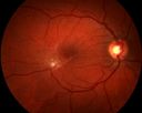
Epiretinal Membrane with Pseudohole1030 views
|
|

Myopic Macular Degeneration - Dry 1029 views55-year-old woman has myopic macular degeneration in both eyes with vision and an Amsler grid change starting a few days ago in the left eye.
VISUAL ACUITY: OD: 20/60. PH: 20/25. OS: 20/200. PH: 20/30.
|
|
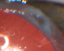
Propionibacterium acnes endophthalmitis with capsular plaque and uveitis1029 views61 year old man with inflammation after cataract surgery who ultimately needed removal of intraocular lens and capsule to quiet eye.
|
|

Ghost Cell Glaucoma1029 viewsPost cataract surgery, patient presents with ghost cell in the anterior chamber after hemorrhage cleared. A small hyphema still is visible in the anterior chamber with a whitish ball appearance.
|
|

PXE with Angioid Streaks1028 viewsPXE with angioid streaks OS (12/09/2011)
|
|

Macroaneurysm FP1026 viewsMacroaneurysm with Subretinal Hemorrhage and ME in OS of an Elderly Female
|
|
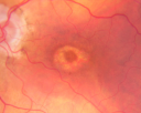
Plaquenil Toxicity - Bulls Eye Maculopathy1025 views70-year-old woman with systemic Lupus erythematosus and clotting problems. She was on the Plaquenil for about eight years and then off the Plaquenil for the last eight years because she developed macular toxicity. Although her vision was hazy, it was stable. Recent deceased vision left eye: OD 20/60, OS 20/100. IOP: OD 18, OS 19.
|
|

Regressed Proliferative Diabetic Retinopathy with Recurrent Vitreous Hemorrhage from Vitreoretinal Traction over a Major Retinal Vessel (Vein)1025 views56-year-old woman has had recurrent vitreous hemorrhage in the left eye from vitreoretinal traction. Diabetes Mellitus for 20 years and excellent response to vitrectomy in the right eye about 1 year ago for recurrent vitreous hemorrhage from traction over a major retinal vessel.
VISUAL ACUITY: Vision OS is 20/200
|
|

1023 views
|
|

Best Disease, Vitelliform Macular Dystrophy - 6 year old child1021 viewsVISUAL ACUITY: Vision OD is 20/30, OS is 20/100.
|
|

DUSN - Diffuse Unilateral Subacute Neuroretinitis - Nematode1021 views
|
|

Myopic Degeneration with RPE Loss1021 viewsPatient comes in for follow up for glaucoma. VA is 20/30 in both eyes. Fundus photography shows the RPE loss due to myopic degeneration, nasally, inferiority in the left eye.
|
|

Acute Zonal Occult Outer Retinopathy1020 views45-year-old man noticed a few years ago peripheral vision blurriness in the left eye and also some central vision loss. Previous to that, as far as he knows, the two eyes were okay.
VISUAL ACUITY: OD: 20/20; OS: 20/40.
|
|

Patterned Pigment Dystrophy of the Fovea 79 YO Woman VA 20/20 Right 20/30 Left1020 views
|
|
| 17559 files on 176 page(s) |
 |
 |
 |
 |
6 |  |
 |
 |
 |
 |
|