|
|

Patterned Pigment Dystrophy of the Fovea 79 YO Woman VA 20/20 Right 20/30 Left567 views
|
|

Birdshot Chorioretinitis - Retinal Vasculitis - HLA A29 Positive 567 views64-year-old woman three or four year’s fluctuations in her vision. She mostly notices them in bright light her vision glares out and she has difficulty seeing. PMHx: malaria in 1948 that was treated. She was told at one point she could possibly have had Familial Mediterranean fever.
She does intermittently get rashes around her arms. She has arthritis in her knees and ankles.
VISUAL ACUITY: OD 20/40, OS 20/50 - HLA - A29 Positive
|
|

Acute Annular Outer Retinopathy for 3 Months - 25 YO Female Spectral Domain Optical Coherence Tomgraphy567 views25-year-old female who, about three months ago, put on her sunglasses and noticed a smudge in the vision and at that point she noticed the right eye was hazy. She had a cold a week or two before the vision loss, but she didn’t have any fever. She has no focal neurological problems in her past history. VA 20/20 OU
|
|

Pigmented Choroidal Mass Right Eye - Melanoma vs. Nevus vs. Unilateral Melanocytic Proliferation - 4 Month Follow-Up Images 567 views71-year-old man his vision 4 month follow-up. Vision is 20/60 and patient sees flashing lights. Lesion has grown slightly in the superior half of the macula.
|
|

Retinal Tear treated with Laser 567 views
|
|

Combined Artery and Vein Occlusion567 viewsPatient comes in with decreased vision in both eyes. VA 20/200-OD, 20/60-OS. Fundus exam shows great amount of macular edema due to artery and vein occlusions. There is some neovascularization on the optic nerve in the right eye. Patient was treated with Eylea Injection in the left eye today and will return for Eylea Injection in the right eye.
|
|

Central Serous Retinopathy 25 year old professional baseball player - Atypical - Choroidal Hypo-perfusion?567 views
|
|

FA showing NVE, NVD, non- perfusion left eye inferonasally with neovascularization at the margin of the non-perfusion567 views
|
|
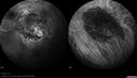
Medium Choroidal Melanoma Left Eye 567 views
|
|

567 views38-year old male presents with decreased vision in the left eye. VA was 20/20-right eye and 20/70-left eye with pinhole no improvement. Fundus exam shows exudates near the macula and also exudates with small hemorrhages, superior nasally in the left eye.
|
|

Chronic Central Serous Retinopathy - Fluorescein Angiogram567 views
|
|

Proliferative Diabetic Retinopathy - Moderate - mild NVD with NVE 567 viewsFA shows leakage from neovascularization and dark areas in periphery from non-perfusion
|
|

Ahmed Tube Placement with Pericardial Patch567 viewsPatient with elevated pressures underwent Ahmed valve placement in the left eye. VA was hand motion. Slit lamp shows a quarter of hyphema in the anterior chamber with Ahmed tube placed temporal at 2-o'clock pressing on the iris. Air bubble is also visible in the anterior chamber in the left eye.
|
|

Macular Star, Toxoplasmosis567 viewsYoung male with visual disturbance in the right eye. Fundus photo shows swelling around the optic nerve with macular star visible.
|
|

Bilateral Papilledema 567 viewsYoung male presents with decreased vision in both eyes. Fundus photos reveal swollen optic nerves in both eyes.
|
|

Macular Hole Left Eye - progression from VMT - Stage II macular hole - Spontaneously Closed Macular Hole567 viewsMacular hole is closing and vision has improved to 20/40
|
|

Sickle Retinopathy - Ultra Wide Field Angiogram - Heidelberg567 viewsLeakage from peripheral fronds of NV - HGB SC disease - 28 year old man with vision loss OS from acute vitreous hemorrhage
|
|

Diffuse outer retinal atrophy in 51 year old woman with 22 years plaquenil (hydroxychlorquine) use567 viewsRetinitis pigmentosa or similar dystrophy vs plaquenil toxicity
|
|

Multifocal Pattern Dystrophy567 views51 year old with normal vision - images look a lot like fundus flavimaculatus
|
|

Optic Nerve Pit Retinal Detachment - SD OCT of Nerve EDI shows Pit567 views9 Year old with morning glory disc who developed nerve pit RD. Raster scans of the optic nerve on EDI setting show the communication between the subretinal space and the fluid around the cerebrospinal fluid surrounding the optic nerve sheath.
|
|
567 views
|
|

Pigmented Paravenous Retinochoroidal Atrophy - Left Eye567 views70 year old woman lost vision OS 20 years ago.
|
|

Outer Retinal Tubulation567 views78 year old man chronic wet AMD - 20/200
|
|

Cat Scratch - Neuroretinitis OD AND retinitis OS - Bartonella henselae positive567 views27 year old female with exposure to cats and prior fever. Reports with recent vision loss right eye. She was treated with doxycycline and within 2 weeks the right eye looks better. Then she skipped all follow-up visits. The right eye presented 20/200 and the left 20/16
|
|

567 views
|
|

Central Retinal Vein Occlusion - New- and old Macular Scar from AMD566 views87-year-old woman with longstaning macular scars and new vision loss left eye for 2 months. OD: 20/200, OS 20/400.
|
|
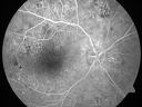
Clinically Significant Diabetic Macular Edema both Eyes with Exudates566 views56-year-old woman with diabetes for twelve years. She has noticed blurring of the vision in the right eye for about six months. OD is 20/70, OS is 20/25.
|
|

Hypertensive Choroidopathy - Pheochromocytoma - Chronic Very High Blood Pressure 49 YO Male566 views49-year-old man chronic very high blood pressure sometimes the blood pressure will go as high as 260/160, sometimes it is 200/120 and sometimes it is 160/80. He has a pheochromocytoma on the adrenal gland and he has had vision loss in the right eye starting between three and four years ago.
VISUAL ACUITY: OD 20/60, OS 20/25.
|
|

Ocular Ishemic Syndrome - Bilateral Carotid Occlusions - OD 20/60 OS 20/200 Enlarged Foveal Avascular Zone and Temporal Non-perfusion566 views75-year-old man had laryngeal cancer treated in 15 year ago with radiation. He recently was told that the carotid circulation is closed off on both sides. He does have a patent vertebral artery, which is supplying him pretty well and in addition to that his cardiac output is compromised by poor mitral valves. His consideration is to what can be done about that in the future.
VISUAL ACUITY: OD 20/60, OS 20/200
|
|

Acute Annular Outer Retinopathy for 3 Months - 25 YO Female FA566 views25-year-old female who, about three months ago, put on her sunglasses and noticed a smudge in the vision and at that point she noticed the right eye was hazy. She had a cold a week or two before the vision loss, but she didn’t have any fever. She has no focal neurological problems in her past history. VA 20/20 OU
|
|
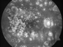
Macular and Peripheral Retinal Scarring from Diabetic Laser Done 25 years previously566 views72-year-old man had macular and peripheral laser done in the past(1980's)(25 years ago). His vision, although limited, seems about stable to him. He is diabetic for 30 years. OD is 20/160, OS is 20/160.
|
|

Angioid Streaks - Peau D'Orange - Pseudoxanthoma Elasticum - Asymptomatic Extrafoveal Choroidal Neovascular Membrane566 views60-year-old woman has pseudoxanthoma elasticum since 2008. Her sister has the same problem and lost vision in both eyes. She had a problem with blood vessel growth in the left eye for which she had injections of Avastin in August, September, and October of 2011 (5 months ago). Subsequent to that, the macula has been dry and you have just been watching her carefully. She is in Florida for a short time but will ultimately be spending nine months out of the year here. Both of her eyes have good vision at
|
|

Lumigan (Bimatoprost) Cystoid Macular Edema - Eye Better 1 month after Stopping Lumigan566 views 82-year-old man had cataract surgery in 1993. He has had glaucoma for seven or eight years for a long time. He is taking Lumigan only in the left eye. About a year ago he started in the right eye and then starting just about a few months ago he has noticed intermittent blurring in the vision in the right eye. Vision 20/25 OD, 20/20 OS
|
|
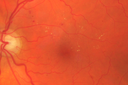
Diabetic Macular Edema - Mild566 views70 year old man with mild edema did well without treatment
|
|

Vasculitis - Retinitis - Uveitis - Vision NLP right eye , 20/50 left eye566 views
|
|

Primary Ocular Lymphoma - 051812 566 views
|
|

Primary Ocular Lymphoma - 051812 566 views
|
|
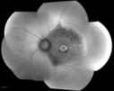
Bullseye Maculopathy without Plaquinel566 viewsFundus photograph reveals Bull's-eye maculopathy without the use of plaquinel. Patients VA is 20/20, Right eye and 20/25, Left eye. There are reported cases of Scleroderma patients with retinal pigment epithelial atrophy. Will return for follow up in 6-months.
|
|

Pseudoxanthoma Elasticum with Angioid Streaks566 viewsYoung female patient who comes in for routine eye exam, VA 20/20 in both eyes. Fundus photos shows a Peu'd orange appearance in the periphery of the retina and angioid streaks around the optic nerve.
|
|

Neuroretinitis - Cat Scratch - Bartonella - Sudden Vision Loss Right Eye - 29 Year Old Female566 views
|
|

Unilateral Acute Idiopathic Maculopathy (UAIM) - VIDEO TAKES TIME TO LOAD - Raster Scan OCT566 viewsVision has improved back to 20/20 without any therapy. 1 month following vision loss
|
|

West Nile Virus Multifocal Choroiditis and diabetic retinopathy566 viewsMultifocal and clustered focal lesions are from west nile virus. The retinopathy is probably diabetic but could also be related to west nile virus
|
|

Syphillitic Optic Neuropathy and Maculopathy566 views46 year old woman with 2 weeks of vision loss OD. VA 20/200 OD; 20/20 OS. there are posterior vitreous cells on OCT in both eyes. ICG, FAF, and FA show an abnormal macula OD. VA improved to 20/30 within a month with treatment with PCN.
|
|
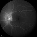
Syphillitic Optic Neuropathy and Maculopathy566 views46 year old woman with 2 weeks of vision loss OD. VA 20/200 OD; 20/20 OS. there are posterior vitreous cells on OCT in both eyes. ICG, FAF, and FA show an abnormal macula OD. VA improved to 20/30 within a month with treatment with PCN.
|
|

Syphillitic Optic Neuropathy and Maculopathy566 views46 year old woman with 2 weeks of vision loss OD. VA 20/200 OD; 20/20 OS. there are posterior vitreous cells on OCT in both eyes. ICG, FAF, and FA show an abnormal macula OD. VA improved to 20/30 within a month with treatment with PCN.
|
|

Cystoid Macular Edema 10 years after Cataract Surgery566 viewsNo overt uveitis but the patient had aching in the eyes and responded to topical anti-inflammatory drops with recurrence of the CME on discontinuation of topical therapy
|
|

Proliferative Diabetic Retinopathy - NVD regressed with PRP laser566 views76 year old diabetic man - Presented one year ago with NVD in the left eye. This regressed with laser but then a year later worsened. Additional PRP was done and the NVD regressed again.
|
|

Diabetic Macular Edema with Exudates566 viewsPatient did not want injection therapy and did well with laser
|
|
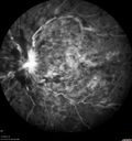
Central Retinal Vein Occlusion with Macular Edema566 views74 year old female The vision in the left eye has gotten worse over the last 3 weeks.
VA OD: Dcc20/25
VA OS: DccCF 3ft
IOP: TP: OD:16 OS:14
Vision improved to 20/40 in 3 months with monthly Aflibercept
|
|
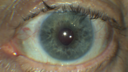
Fuch's Heterochromic Iridocyclitis - Iris Photos566 views71 year old man with inflammation in the left eye for over 50 years. The left eye got a tube shunt 15 years ago. Vision is 20/32 OD, 20/40 OS
|
|

Chronic Pseudophakic Cystoid Macular Edema in a Patient with Diabetes565 views74-year-old woman has chronic cystoid macular edema in the left eye. She has had vision loss intermittently in that eye for 2 years, the right eye, unfortunately, lost vision from a central retinal vein occlusion in 6 years ago.
|
|

Angioid Streaks - New Extrafoveal Choroidal Neovascular Membrane Right Eye565 views61-year-old woman twelve days ago, saw a spot to the right of her central vision. OD is 20/25, OS is 20/25
|
|

Hypertensive Choroidopathy - Pheochromocytoma - Chronic Very High Blood Pressure 49 YO Male565 views49-year-old man chronic very high blood pressure sometimes the blood pressure will go as high as 260/160, sometimes it is 200/120 and sometimes it is 160/80. He has a pheochromocytoma on the adrenal gland and he has had vision loss in the right eye starting between three and four years ago.
VISUAL ACUITY: OD 20/60, OS 20/25.
|
|

Macular Fold - 40 years post retinal detachment repair - Glaucoma 565 views79-year-old woman has had a long-standing macular fold in the right eye associated with retinal detachment repair in 1970. (40 years ago) She has a macular scar in that eye. She has optic atrophy in both eyes from glaucoma. Vision OD is hand motion, OS is 20/50
|
|
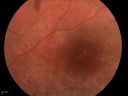
DUSN - Diffuse Unilateral Subacute Neuroretinitis - Nematode565 views
|
|

Myopic Traction Maculopathy - Vitreoretinal Traction - Possible Occult Neovascular Membrane565 views58-year-old woman was seen in the office on June 30, 2011. In 2002, her vision was 20/30. Subsequent to that she had her left eye made her distance eye, which was dominate. She had LASIK surgery on the right eye to make that her near eye and she was getting along well until about a year ago when she noticed substantial vision loss in the left eye only. VISUAL ACUITY: The near eye is 20/200, but she reads J2. OS 20/100- and J16.
|
|

Myopic Traction Maculopathy - Vitreoretinal Traction - Possible Occult Neovascular Membrane565 views58-year-old woman was seen in the office on June 30, 2011. In 2002, her vision was 20/30. Subsequent to that she had her left eye made her distance eye, which was dominate. She had LASIK surgery on the right eye to make that her near eye and she was getting along well until about a year ago when she noticed substantial vision loss in the left eye only. VISUAL ACUITY: The near eye is 20/200, but she reads J2. OS 20/100- and J16.
|
|

Polypoidal Choroidal Vasculopathy Right Eye - Disciform Scar Left Eye565 views84-year-old woman has polypoidal choroidal vasculopathy in both eyes with macular degeneration. She has macular scars in both eyes and she remains with remarkably good vision in the right eye despite the scarring.
VISUAL ACUITY: OD 20/30, OS 1/200
Avastin Rx OD
|
|

Acute Annular Outer Retinopathy for 3 Months - 25 YO Female Fundus Photograph565 views25-year-old female who, about three months ago, put on her sunglasses and noticed a smudge in the vision and at that point she noticed the right eye was hazy. She had a cold a week or two before the vision loss, but she didn’t have any fever. She has no focal neurological problems in her past history. VA 20/20 OU
|
|

Malignany Hypertension - BP 250/130 - Vision 1/200 in each eye - Diabetic565 views68-year-old woman poor vision in both eyes for the last few years. She is diabetic since 1989. Her blood pressure has been very high. She was in the hospital Friday. Her blood pressure during that visit was 250/130 and her blood pressure in your office she reports was somewhere around the same. I just checked her blood pressure at the beginning of my visit and it was 200/120 in her right arm sitting. Her vision is poor enough that she can barely see to walk around. She has also been feeling nauseous
|
|

Pigmented Choroidal Mass Right Eye - Melanoma vs. Nevus vs. Unilateral Melanocytic Proliferation -4 Month Follow-Up Images565 views71-year-old man his vision 4 month follow-up. Vision is 20/60 and patient sees flashing lights. Lesion has grown slightly in the superior half of the macula.
|
|

Non-Ischemic Central Retinal Vein Occlusion in Young Man (30 years) - Many Cotton Wool Spots565 views30-year-old man with a history of a central retinal vein occlusion for which he was first examined on 2 months ago and his vision then was 4/200. He responded nicely to two Avastin treatments. OD is 20/20, OS is 20/60
|
|

Chronic Central Serous Retinopathy - Fluorescein Angiogram565 views
|
|

Proliferative Diabetic Retinopathy - Moderate - mild NVD with NVE 565 viewsFA shows leakage from neovascularization and dark areas in periphery from non-perfusion
|
|

Ahmed Tube Placement with Pericardial Patch565 viewsPatient with elevated pressures underwent Ahmed valve placement in the left eye. VA was hand motion. Slit lamp shows a quarter of hyphema in the anterior chamber with Ahmed tube placed temporal at 2-o'clock pressing on the iris. Air bubble is also visible in the anterior chamber in the left eye.
|
|

Pseudoxanthoma Elasticum with Angioid Streaks565 viewsYoung female patient who comes in for routine eye exam, VA 20/20 in both eyes. Fundus photos shows a Peu'd orange appearance in the periphery of the retina and angioid streaks around the optic nerve.
|
|

Unilateral Acute Idiopathic Maculopathy (UAIM)565 views41 year old with serous retinal detachment OS and sudden vision loss. He had a viral prodrome. Coxscackie IgG was high but no acute infection was documented. Vision improved in about 2 months without therapy. - Right eye has spot nasal to the optic nerve
|
|

Recurrent MEWDS Left Eye - Teenage Girl565 viewsPatient presented in 2014 (first images) 2 months following vision loss left eye. Vision went from 20/60 - 20/20. OCT showed outer retinal irregularities which normalized. Then 3 years later in 2017 she had full blown MEWDS. She presented acutely and all the images are typical. Both episodes where in the same time of year, about 3 months after her flu shot for sports.
|
|

Branch Retinal Vein Occlusion (BRVO) no macular edema - cotton wool spot565 views85 year old man with asymptomatic BRVO - his vision stayed great without needing any treatment.
|
|

Anemic Retinopathy565 viewsMyelodysplasia - HgB 8 and very low platelet count
|
|

Central Retinal Vein Occlusion - No macular edema - Pre-foveal opercula565 views62 year old female with 20/20 vision. She had been seeing a spot out of the left eye for 4 years and then noticed splotches in her vision. She never devleoped edema or NV
|
|

Diabetic Macular Edema - One eye treated with Eylea and 2 weeks later both were improved565 views68 year old female with diabetic retinopathy and macular edema
VA OD: sc20/40 PH20/32
VA OS: sc20/40 PH20/32
IOP: TP: OD:14 OS:14 Time:12:45
|
|

Stellate Non-heredtiary Idiopathic Foveomacular Retinoschisis (SNIFR)565 views77 year old man who is healthy and 6'6" tall with 20/40 vision and no complaints. OCT shows diffuse retinoschisis.
|
|
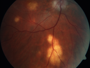
Multifocal Choroiditis - Pneumocystis - aspergillis - cryptococcus565 views31 year old male with anaplastic astrocytoma on chemotherapy with mild vision loss in the right eye. LP did not reveal organism. He was placed on a trial of anti-fungal medications and lost to follow-up
|
|

Retinal fold following removal of severe macular pucker565 views75 year old man had macula on inferior retinal detachment repair. He then developed a 5/200 dense macular pucker. After the pucker was removed the vision returned to 20/40 but there was a persistent superior retinal fold that looks a little like a snake.
|
|

Choroidal Melanoma Medium564 views33-year-old man with a history of decreased in the right eye. He had blurring in the eye about a year ago. He reports that it improved spontaneously until it was “nearly resolvedâ€. He said he had some persistent distortion, but that over the past couple of weeks he has noticed this dark spot recurring in the right eye, the left eye is unaffected. He does not have any flashes, floaters or change in his peripheral visual acuity. 20/80 OD, 20/16 OS. No improvement with pinhole. IOP: 12 OD, 11 OS.
POSTERIOR SEGMENT EXAMINATION: Shows a clear view with no PVD in either eye. C/D ratio is 0.2 OU. Large retinal vessels are unremarkable. The macula of the right eye has a brown choroidal lesion with pigment overlying the surface and orange spots along its lateral surface. The left macula is normal.
B-SCAN: B-scan ecography of the ultrasound of the right eye shows a mass about 5 mm high with a moderate to low internal reflectivity on A-scan echogram. There is serous fluid inferiorly on the echogram
|
|
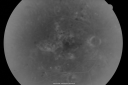
BDR and Diabetic Macular Edema with Fresh Laser Right Eye564 views60-year old man OD 20/60, OS 20/50 worsening vision both eyes
|
|

564 views49-year-old woman has central retinal vein occlusion right eye with fluctuating vision for a few month now with OD 20/100. IOP: OD 20
|
|

Patterned Pigment Dystrophy of the Fovea 79 YO Woman VA 20/20 Right 20/30 Left564 views
|
|

80 Year old man with 3 day history of vision loss right eye. Vision 4/200.564 views
|
|

Pigmented Maculae - Mother and Daughter - Normal Visual Acuity564 views12-year-old had a dilated macular exam and the macula look abnormal. Interestingly, her mother, who is with her at this visit, was told about a year ago that her maculae are abnormal too. Her mother’s vision is fine. In the family tree, there are no known retinal dystrophies. They are both healthy.
VISUAL ACUITY: Her vision is 20/20 in both eyes.
|
|

Acute Central Serous Retinopathy (Chorioretinopathy) Right Eye564 views45-year-old man decreased vision in the right eye starting about two days ago. VISUAL ACUITY: Vision OD is 20/60, OS is 20/20.
|
|

564 views63-year-old man diabetic for twenty two years. His central vision is fine.
VISUAL ACUITY: OD 20/25, OS 20/25
|
|

Acute Central Serous Retinopathy - CSR - Steroid Induced (probably) - Good Vision - FA Affected Eye564 views42-year-old man was seen in the office on October 5, 2011. He had noticed starting in August after a course of antibiotic and steroids, that he developed new spots in his vision in the right eye. He may have had an episode like this sometime in the past. He did take steroids a few years ago and his vision did change at that time, but then returned.
VISUAL ACUITY: OD 20/32, OS 20/32. The OCT scan of the right eye confirms subretinal fluid.
|
|

Uveitis - Retinitis - Vasculitis - possibly Syphilitis564 viewsVisit 1: 12-06-12
74-year-old man 12-3-12 visit. 2 months ago noticed vision loss, first in the right eye than the left. He is getting tired easily. He can’t walk for a long distance. He has lost five pounds within the last month. He also had a severe itchy episode, but not quite a rash a few days ago, which went away after he took a shower. FTA-ABS is positive but a spinal tap, which was negative. OD 1/200, OS 20/63.
|
|

BDR right eye - previous laser564 views
|
|

Chronic Central Serous Retinopathy - IR564 views
|
|

Choroidal Neovascular Membrane - Multifocal Scars - PIC vs MFC vs OHS 564 views
|
|

Embolic CRAO Mid564 views
|
|

Acute AION OS - Mild disc pallor OD564 viewsSudden vision loss OS x 1 week with optic nerve swelling. Evidence of optic atrophy in right eye.
|
|

Posterior Uveal Effusion564 views
|
|

Hydroxychlorquine (Plaquenil) Toxicity - Taiwanese 56 year old female (Asian)564 views56 year old female on 400 mg plaquenil for 22 years presents with vision loss. VA 20/50 both eyes. Plaquenil was stopped. The vision in the left eye continued to deteriorate to 3/200. 5'2" 127 lb (6.6 mg/kg body weight)
|
|

Macular Telangiectasia and dry AMD - Simulating wet AMD564 viewsThis patient was told different things by different retina specialists. Her case is complex because she has OCT findings of Macular Telangiectasis and dry AMD. This makes it look like she has wet AMD. Her vision is stable.
|
|

Coats' Disease - 14 year old - Asymptomatic564 viewsPresented with macular exudates. Temporal macroaneurysms responded to laser. Ultra-wide field FA shows nonperfusion
|
|

Asymptomatic wet AMD - Subfoveal CNM both eyes564 views82 year old woman - 20/25 OU. Images show occult subfoveal CNVM both eyes best seen on late ICG. The left eye also has probable polypoidal choroidal vasculopathy. No treatment was recommended on the first visit - follow-up at 1 month.
|
|

Cystoid Macular Edema following Cataract surgery and vitrectomy for floaters564 views73 year old woman who had decreased vision 4 months following combined vitrectomy and cataract surgery. VA 20/63 improved in 1 month to 20/25 with ketorolac and prednisolone drops.
|
|

Diabetic Macular Edema with Exudates564 viewsPatient did not want injection therapy and did well with laser
|
|

PDR - Mild Vitreous Hemorrhage and Good Vision563 views58-year-old woman has diabetic retinopathy in both eyes with neovascularization of the optic nerve, worse in the right eye than the left eye. She has had panretinal laser in both eyes and vitreous hemorrhage in the left eye. Her vision, since I saw her last in June, is about the same.
VISUAL ACUITY: Vision OD is 20/25, OS is 20/20.
|
|

Central Retinal Artery Occlusion Left Eye 2 Weeks Old - Still Non-Perfusion563 views85-year-old woman who had sudden severevision loss in the left eye two weeks ago. she can see a little more centrally and much more in the periphery, since about the last four days.
VISUAL ACUITY: OD 20/30, OS: Hand motion
|
|
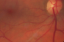
Cotton Wool Spots now Gone in Both Eyes563 views60-year-old man was sick in the hospital in 3 month ago with streptococcal pneumonia. He was in the cardiac intensive care unit and at one point he was in critical condition. He has no visual complaints. OD 20/20, OS 20/25. Cotton Wool Spots now GONE
|
|
| 18117 files on 182 page(s) |
 |
|
|
|
|
61 | |
|
|
|
|


