|
|

Nonproliferative Diabetic Retinopathy8295 views65-year old female with diabetes. Has had cataract surgery in the left eye with VA 20/25. She has had laser in the past. Fundus examination shows microaneurysms with retinal hemorrahages and exudates in the left eye.
|
|

Nonproliferative Diabetic Retinopathy7635 views65-year old female with diabetes. Has had cataract surgery in the left eye with VA 20/25. She has had laser in the past. Fundus examination shows microaneurysms with retinal hemorrahages and exudates in the left eye.
|
|

Nonproliferative Diabetic Retinopathy6749 views65-year old female with diabetes. Has had cataract surgery in the left eye with VA 20/25. She has had laser in the past. Fundus examination shows microaneurysms with retinal hemorrahages and exudates in the left eye.
|
|

Optic disc swelling6119 views56 year old woman
|
|

Nonproliferative Diabetic Retinopathy5896 views65-year old female with diabetes. Has had cataract surgery in the left eye with VA 20/25. She has had laser in the past. Fundus examination shows microaneurysms with retinal hemorrahages and exudates in the left eye.
|
|

Nonproliferative Diabetic Retinopathy5824 views65-year old female with diabetes. Has had cataract surgery in the left eye with VA 20/25. She has had laser in the past. Fundus examination shows microaneurysms with retinal hemorrahages and exudates in the left eye.
|
|

Amelanotic - Light Colored CHRPE - Congenital Hypertrophy of the Pigment Epithelium5160 views56-year-old woman nov vision problems. OD is 20/20, OS is 20/16
|
|

Juvenile Onset Diabetic - Proliferative Diabetic retinopathy with NVE and NVD4621 viewsProliferative Diabetic Retinopathy with Neovascularization of the Disc and Neovascularization Elsewhere.
James L. Perron, C.R.A.
|
|
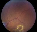
Nonproliferative Diabetic Retinopathy4483 views65-year old female with diabetes. Has had cataract surgery in the left eye with VA 20/25. She has had laser in the past. Fundus examination shows microaneurysms with retinal hemorrahages and exudates in the left eye.
|
|

PVD with Weiss Ring /Optic Nerve in Background4444 views
|
|

Plaquenil Toxicity both Eyes - Partial Bull's Eye - Discontinued 6 Years ago SD-OCT (Spectral domain optical coherence tomography)4276 views82-year-old woman was on Plaquenil from 1976 from 2005, 200 mg a day. It was discontinued because of abnormal visual fields 6 years ago. OD 20/32, OS 20/40
|
|

Purtscher's Retinopathy - Motor Vehicle Accident4181 viewsNerve Fiber Layer Inflammation and retinal hemorrhages due to severe trauma. Patient was a teenage boy that got into a car accident.
|
|

Type 1 Retinal Arteriovenous Malformation4104 views42 year old female VA 20/32 OD, 20/20 OS MRI was negative.
|
|

CHRPE - Gardner Syndrome - FAMILIAL COLORECTAL POLYPOSIS - 11 year old female4074 views11-year-old child with CHRPE OS has six undescended adult teeth and also she has had a cyst removed from her arm pit and she has three more in her arm pit and one on her face and she is seeing a dermatologist for that in the near future. The mother reports no health problems, however, the patient’s grandmother does have a history of intestinal polyps, as does the patient’s grandmother’s brothers son.
|
|

Wyburn Mason Syndrome4041 views28 year old female, visual acuity OS 20/200
|
|
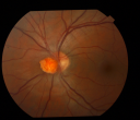
Optic Nerve Tumor3839 views21 year old male: Presents with a history of Von Hippel-Lindau Syndrome, diagnosed in 2005 affecting his left Optic Nerve, Brain, Spine, Kidney and Pancreas. He has undergone Laser for Retinal Hemangioma measuring 2/3-4/5DD -vs_ 1/3DD when originally diagnosed. However his visual acuity remains good 20/25. Patient has also undergone Neurosurgery in 2005 and Spinal cord in 2006. Recent MRI of spinal cord hemangioma showed stable tumors.
|
|

Plaquenil Toxicity both Eyes - Partial Bull's Eye - Discontinued 6 Years ago SD-OCT (Spectral domain optical coherence tomography)3746 views82-year-old woman was on Plaquenil from 1976 from 2005, 200 mg a day. It was discontinued because of abnormal visual fields 6 years ago. OD 20/32, OS 20/40
|
|

Sarcoidosis Multifocal Choroiditis3706 views66 year old man with dense cataracts and recurrent uveitis. Images show multifocal choroidal granulomas from sarcoidosis more in the right eye than the left eye.
|
|

Occult Metallic Intraocular Foreign Body - WINNER JANUARY 2011 - BEST IMAGE3617 viewsFundus photograph of a 28 year old male who presented with intermittent, painless, blurred vision of his left eye secondary to an occult metallic intraocular foreign body with old vitreous hemorrhage. He later admitted to feeling a mild gritty sensation of his affected eye while working with a circular saw the previous year. He was then misdiagnosed as having a subconjunctival hemorrhage from the emergency physician. He later underwent successful pars plana vitrectomy and foreign body removal.
|
|

Plaquenil Toxicity both Eyes - Partial Bull's Eye - Discontinued 6 Years ago SD-OCT (Spectral domain optical coherence tomography)3608 views82-year-old woman was on Plaquenil from 1976 from 2005, 200 mg a day. It was discontinued because of abnormal visual fields 6 years ago. OD 20/32, OS 20/40
|
|

Peripheral Bullous Retinoschisis Spectral Domain Optical Coherence Tomography (SD-OCT)3261 views64-year-old man with peripheral retinal problem noted during comprehensive eye examination. OD 20/32, OS 20/25
|
|

Proliferative Diabetic Retinopathy with Macular Hole3154 viewsPatitent with diabetes diagnosed 12 years ago and low visual acuity.
|
|

Reticular Macular Disease - Reticular Pseudodrusen and Soft Macular Drusen - 57 Year Old Woman3076 views57-year-old woman was seen in the office on 2/22/2011. She has macular drusen in both eyes. I saw her back in 2006. Since then she does notice slight increased distortion. OD is 20/25, OS is 20/25
|
|
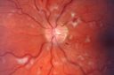
Non-Ischemic Central Retinal Vein Occlusion in Young Man (30 years) - Many Cotton Wool Spots3030 views30-year-old man with a history of a central retinal vein occlusion for which he was first examined on 2 months ago and his vision then was 4/200. He responded nicely to two Avastin treatments. OD is 20/20, OS is 20/60
|
|

Macular fold after PPV for RD3030 viewsA 53-year-old man with a macula-off RD underwent left eye pars plana vitrectomy with air-fluid exchange, laser retinopexy, and injection of 14% SF6 gas. There were 4 retinal breaks between 10 o’clock and 12 o’clock including one large tear. He was compliant with facedown positioning. On the 8th post-operative day, a retinal fold along the 4-10 o’clock meridian was seen coursing through his central macula. He underwent repeat PPV but multiple attempts to lift and flatten the retina were unsuccessful.
|
|

CAVERNOUS HEMONGIOMA2918 views28 year old male w/ 20/200 vision at time of exam. Patient c/o poor vision since childhood. No significant medical history or family medical history. A problem was only noted when patient enlisted in the Army.
|
|

Nonproliferative Diabetic Retinopathy2894 views65-year old female with diabetes. Has had cataract surgery in the left eye with VA 20/25. She has had laser in the past. Fundus examination shows microaneurysms with retinal hemorrahages and exudates in the left eye.
|
|

Combined hamartoma of the retina and RPE2838 views16 year old female, diagnosed with a combined hamartoma of the retina and the retinal pigment epithelium.
|
|

Prominent Posterior Hyaloid with Background Diabetic Retinopathy2833 viewsDiabetic patient comes in for follow up for her Background Diabetic Retinopathy and glaucoma. VA is 20/30. left eye. Fundus exam shows posterior Hyaloid with hemorrhage inferiorily.
|
|

Plaquenil Toxicity both Eyes - Partial Bull's Eye - Discontinued 6 Years ago SD-OCT (Spectral domain optical coherence tomography)2812 views82-year-old woman was on Plaquenil from 1976 from 2005, 200 mg a day. It was discontinued because of abnormal visual fields 6 years ago. OD 20/32, OS 20/40
|
|

Amelanotic - Light Colored CHRPE - Congenital Hypertrophy of the Pigment Epithelium2791 views56-year-old woman nov vision problems. OD is 20/20, OS is 20/16
|
|

Acute Macula Neuroretinopathy2788 views39yr old male: Presents with Inferior Temporal Scotoma in his left eye, x 10 days with no change in shape or size, Visual acuity 20/25.
Most common sysptoms are described as sudden onset of one or more paracentral scotomas. {with the tip pointing toward the Fovea} without any other visual symptoms. Currently no treatment recommended.
|
|

Asymptomatic Hollenhorst Plaque - Cholesterol Embolis2667 views71-year-old woman is not noticing any vision change. She does take pressure-lowering drops for glaucoma, both Timolol and Xalatan.
VISUAL ACUITY: Vision OD is 20/30, OS is 20/16
|
|

Anterior Ischemic Optic Neuropathy - Non-Arteritic - AION2652 views72-year-old woman sudden vision loss in the right eye about four days ago. She also has noticed that things are dim out of that eye. She has no headaches. She does feel a little tired. She has no pain in her jaw when she chews and she has no fevers.
OD is 20/80, PH 20/25; OS is 20/25
|
|

Plaquenil Toxicity both Eyes - Partial Bull's Eye - Discontinued 6 Years ago Fundus Autofluorescence2646 views82-year-old woman was on Plaquenil from 1976 from 2005, 200 mg a day. It was discontinued because of abnormal visual fields 6 years ago. OD 20/32, OS 20/40
|
|

Horseshoe Retinal Tear with bridging blood vessel2614 views
|
|

Correctopia - post laser iridoplasty - 20/20 vision2608 views1 month after the iridoplasty, her vision was 20/20. She began to improve on the drive home from the treatment. The laser setting were : green argon 500 microns, 500 mw, 500 msec.
|
|
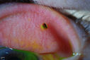
Corneal Abrasion with Foreign Body Present 2603 viewsER patient comes in with corneal abrasion in the left eye which was getting worst. VA 20/25. Slit lamp exam showed corneal abrasion superiorly at 2-o'clock. Flipped eyelid and foreign body appeared on the upper tarsal plate. Foreign body was removed.
|
|

Prominent Posterior Hyaloid with Background Diabetic Retinopathy2508 viewsDiabetic patient comes in for follow up for her Background Diabetic Retinopathy and glaucoma. VA is 20/30. left eye. Fundus exam shows posterior Hyaloid with hemorrhage inferiorily.
|
|
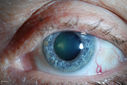
Dilated Conjunctival Blood Vessels2507 viewsPatient comes in for routine eye exam. VA 20/20 in both eyes. Fundus exam was normal. Dilated blood vessels are shown temporal inferiorly in the left eye.
|
|

Retinal Vasculitis due to Lupus (color)2501 viewsSystemic Lupus Erythematosis - Vasculitis
|
|

Serous Choroidal Effusions2473 viewsSerous choroidal effusions following anterior segment surgery. Final visual outcome was 20/20.
|
|

"Jelly Bumps" Soft Contact Lens2417 viewsYoung female patient comes in wearing overnight contact lenses. Complains of irritation in both eyes. Denies sleeping in SCL. Slit lamp exam shows little jelly bumps underneath the contact lens in both eyes. Was issued a new pair and lectured on handling them.
|
|
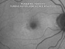
Plaquenil Toxicity both Eyes - Partial Bull's Eye - Discontinued 6 Years ago Fundus Autofluorescence2410 views82-year-old woman was on Plaquenil from 1976 from 2005, 200 mg a day. It was discontinued because of abnormal visual fields 6 years ago. OD 20/32, OS 20/40
|
|

Corneal Abrasion with Foreign Body Present 2409 viewsER patient comes in with corneal abrasion in the left eye which was getting worst. VA 20/25. Slit lamp exam showed corneal abrasion superiorly at 2-o'clock. Flipped eyelid and foreign body appeared on the upper tarsal plate. Foreign body was removed.
|
|

Non-Ischemic Central Retinal Vein Occlusion in Young Man (30 years) - Many Cotton Wool Spots2405 views30-year-old man with a history of a central retinal vein occlusion for which he was first examined on 2 months ago and his vision then was 4/200. He responded nicely to two Avastin treatments. OD is 20/20, OS is 20/60
|
|

OD Albinism with foveal hypoplasia2396 views28 year old female latina with vision of 20/400
|
|

Psuedo-retinitis Pigmentosa - Bone Spicules One Eye - Probably Acute Zonal Occult Outer Retinopathy (AZOOR)2382 views65-year-old woman has pseudoretinitis pigmentosa in the right eye only, most likely from acute zonal occult outer retinopathy. OD 20/25, OS 20/25
|
|

Nonproliferative Diabetic Retinopathy2379 views65-year old female with diabetes. Has had cataract surgery in the left eye with VA 20/25. She has had laser in the past. Fundus examination shows microaneurysms with retinal hemorrahages and exudates in the left eye.
|
|

Dilated Conjunctival Blood Vessels2375 viewsPatient comes in for routine eye exam. VA 20/20 in both eyes. Fundus exam was normal. Dilated blood vessels are shown temporal inferiorly in the left eye.
|
|

Anterior Ischemic Optic Neuropathy - Non-Arteritic - AION2361 views72-year-old woman sudden vision loss in the right eye about four days ago. She also has noticed that things are dim out of that eye. She has no headaches. She does feel a little tired. She has no pain in her jaw when she chews and she has no fevers.
OD is 20/80, PH 20/25; OS is 20/25
|
|

Dilated Conjunctival Blood Vessels2348 viewsPatient comes in for routine eye exam. VA 20/20 in both eyes. Fundus exam was normal. Dilated blood vessels are shown temporal inferiorly in the left eye.
|
|

CAVERNOUS HEMONGIOMA2280 views28 year old male. FA
|
|

Reticular Macular Disease - Reticular Pseudodrusen and Soft Macular Drusen - 57 Year Old Woman2244 views57-year-old woman was seen in the office on 2/22/2011. She has macular drusen in both eyes. I saw her back in 2006. Since then she does notice slight increased distortion. OD is 20/25, OS is 20/25
|
|
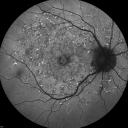
Fundus Flavimaculatus - Stargardt Disease - 20/50 OD 20/200 OS 61 Year old Fundus Autofluorescence od2241 views61-year-old decreasing vision for about the last five years. OD 20/50, OS 20/200.
Pisciform Lesions and Macular Atrophy
|
|

Combined schisis-rhegmatogenous RD2241 viewsMultiple outer layer and inner layer holes
|
|

Choroidal Nevus with Overlying Drusen2222 views48-year-old woman Vision OD is 20/16, OS is 20/20.
|
|
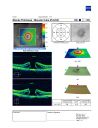
Large mac hole OCT2221 views76 yof with large mac hole
VA: CF at 3ft
chance of spontaneous closure low; surgery option available
|
|
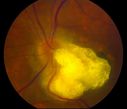
Astrocytic Hamartoma2217 viewsCalcified astrocytic hamartoma of the optic nerve
|
|
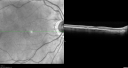
Plaquenil Toxicity both Eyes - Partial Bull's Eye - Discontinued 6 Years ago SD-OCT (Spectral domain optical coherence tomography)2213 views82-year-old woman was on Plaquenil from 1976 from 2005, 200 mg a day. It was discontinued because of abnormal visual fields 6 years ago. OD 20/32, OS 20/40
|
|

Retinal Flap Tear - Horseshoe - Cystic Retinal Tuft on Flap2186 views73-year-old woman who noticed two days ago she was bending over and had a sudden “ribbon†in her
vision which looked to her like blood. VISUAL ACUITY: Vision OD is 20/25, OS is 20/20.
|
|

Diabetic Macular Edema and Hypertensive Retinopathy - Circinate Exudate (Ring Exudate) 2151 views55-year-old woman diabetic for fifteen years and high blood pressure, especially over the last few months. She has had problems with nosebleeds, headaches, and there has been some difficulty bringing her blood pressure down. She said now the blood pressure is under control but it was running 220/120 mmHg for some time. Her vision in the right eye has been poor for four weeks with a spot in the central vision and both eyes have been blurred.
VISUAL ACUITY: Vision OD is 20/160, OS is 20/60
|
|

Malignant Hypertension - Cotton Wool Spots - Elschnig Spots - Optic Nerve Edema 2150 viewsElschnig Spots
|
|

Uveitis - Iris Nodule - Koeppe Nodule2118 views29-year-old woman has had problems with uveitis in the right eye for about the last two years. Her symptoms are usually floaters. Occasionally the eye gets red, it has not been very uncomfortable, and it has always been in the right eye. Has had Iris Nodule like one in photo in left eye and Right eye. They clear with or without steroids after about a month.
|
|

CHRPE - Gardner Syndrome - FAMILIAL COLORECTAL POLYPOSIS - 11 year old female2112 views11-year-old child with CHRPE OS has six undescended adult teeth and also she has had a cyst removed from her arm pit and she has three more in her arm pit and one on her face and she is seeing a dermatologist for that in the near future. The mother reports no health problems, however, the patient’s grandmother does have a history of intestinal polyps, as does the patient’s grandmother’s brothers son.
|
|

Fundus Flavimaculatus - Stargardt Disease - 20/50 OD 20/200 OS 61 Year old - SD - OCT OS2099 views61-year-old decreasing vision for about the last five years. OD 20/50, OS 20/200.
Pisciform Lesions and Macular Atrophy
|
|

Wyburn-Mason Syndrome 2099 views28 year old female, diagnosed with Wyburn-Mason Syndrome at age 7. At time of exam, vision in the left eye was 4/200.
|
|

Myelinated Nerve Fiber Layer Right Eye (white area)2097 views62-year-old man myelinated nerve fiber layer in the right eye. OD 20/25, OS 20/30.
|
|

Peripheral Bullous Retinoschisis Fundus Photo (superotemporal)2089 views64-year-old man with peripheral retinal problem noted during comprehensive eye examination. OD 20/32, OS 20/25
|
|

ANGIOID STREAKS2078 viewsA 29 year old woman was referred to the ophthalmologist for an eye exam since the dermatologist had noted “chicken-skin†papules in the neck skin. PAD showed for pseudoxathoma elasticum typical pathology. The eye exam disclosed angioid streaks, asymptomatic so far.
Magnus Gjötterberg MD
International membrer 1440
S:t Erik Eye Hospital
SE-11282
Stockholm
Sweden
magnusgjotterberg@hem.utfors.se
magnus.gjotterberg@sankterik.se.
Photographer/technician
Malin Langhals
|
|

Conjunctival Cyst2064 viewsPatient comes in with mechanical irritation in the right eye. No foreign body was found. Slit lamp photo shows conjunctival cyst was found temporal in the right eye. Patient will proceed with steroid/antibiotic drops and will stay out of contact lenses.
|
|

Amelanotic Choroidal Nevus 2062 views45-year-old man OD 20/20, OS 20/20. 5x5 mm flat, yellow choroidal lesion just inferonasal to the optic nerve, two disc-diameters from the nerve with scalloped edges. It looks like a confluent drusen, but it is not elevated.
|
|

Bilateral Diffuse Uveal Melanocytic Proliferation - BDUMP - Paraneoplastic Syndrome2059 views80-year-old man vision loss for one year. He died about one year after these photos from Metastatic Poorly Differentiated Large Cell Carcinoma of unknown primary. He was a smoker.
|
|

Reticular Macular Disease - Reticular Pseudodrusen and Soft Macular Drusen - 57 Year Old Woman2048 views57-year-old woman was seen in the office on 2/22/2011. She has macular drusen in both eyes. I saw her back in 2006. Since then she does notice slight increased distortion. OD is 20/25, OS is 20/25
|
|
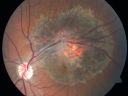
Retinal Pigment Epithelial Dysgenesis2045 views
|
|

Malattia leventinese2042 viewsBasal laminar drusen in a perifoveal radial distribution are noted in this rare condition. Inheritance is AD with variable expressivity but full penetrance.
|
|

Reticular Macular Disease (Pseudo-drusen) Both Eyes - Wet AMD OS - Dry AMD OD SD-OCT2028 views84-year-old woman has wet age-related macular degeneration in the left eye and dry macular degeneration in the right eye. She takes the eye vitamins and her vision is stable since she was treated three months ago with Avastin. OD 20/50, OS 20/32
|
|
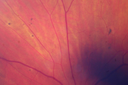
Malignant Hypertension - Cotton Wool Spots - Elschnig Spots - Optic Nerve Edema 2026 viewsElschnig Spots
|
|

CHRPE lesion in the left eye - Irregular pigmentation2012 views63 year old female with normal vision and CHRPE lesion in the right eye.
|
|

Red Free Hemangioma left eye2011 views
|
|

Retinoblastoma2008 viewsYoung male who has a retinal tumor inferior with a calcium core in the left eye. This is a regressed retinoblastoma.
|
|

Anterior Ischemic Optic Neuropathy - Non-Arteritic - AION2002 views72-year-old woman sudden vision loss in the right eye about four days ago. She also has noticed that things are dim out of that eye. She has no headaches. She does feel a little tired. She has no pain in her jaw when she chews and she has no fevers.
OD is 20/80, PH 20/25; OS is 20/25
|
|

Correctopia - post laser iridoplasty - 20/20 vision2002 views1 month after the iridoplasty, her vision was 20/20. She began to improve on the drive home from the treatment. The laser setting were : green argon 500 microns, 500 mw, 500 msec.
|
|

Plaquenil Toxicity both Eyes - Partial Bull's Eye - Discontinued 6 Years ago SD-OCT (Spectral domain optical coherence tomography)1995 views82-year-old woman was on Plaquenil from 1976 from 2005, 200 mg a day. It was discontinued because of abnormal visual fields 6 years ago. OD 20/32, OS 20/40
|
|
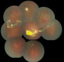
coats disease; exudative retinitis; retinal telangiectasis; leber multiple miliary aneurysm disease1994 viewsclassic coats
|
|

Pigment migration in dry age-related macular degeneration1993 views80 year old female. Dry AMD with GA in the left eye and pigment migration visible on OCT scan.
VA 20/40 OD, 20/160 OS
|
|

Plaquenil Toxicity both Eyes - Partial Bull's Eye - Discontinued 6 Years ago Fundus Autofluorescence1980 views82-year-old woman was on Plaquenil from 1976 from 2005, 200 mg a day. It was discontinued because of abnormal visual fields 6 years ago. OD 20/32, OS 20/40
|
|

Recurrent Macula Off Retinal Detachment - Corrugated Shallow Appearance1977 views62-year-old man shadow a week ago.
VISUAL ACUITY: Vision OS is 20/60. RECURRENT RETINAL DETACHMENT
|
|

Conjunctival Cyst1975 viewsPatient comes in with mechanical irritation in the right eye. No foreign body was found. Slit lamp photo shows conjunctival cyst was found temporal in the right eye. Patient will proceed with steroid/antibiotic drops and will stay out of contact lenses.
|
|
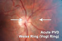
Weiss Ring -Vogt Ring - Posterior Vitreous Detachment1974 viewsChronic Floater - 10 years
|
|

Ectopia Lentis - Marfan's Syndrome - Intermittent Pupillary Block Glaucoma Right Eye1964 views41-year-old woman decreased vision right eye and intermittent pupillary block glaucoma for 6 months from a dislocated natural lens in the eye, probably associated with Marfan syndrome.Vision OD is 20/160, PH 20/40; OS is 20/30, PH 20/25
|
|

Sturge-Weber Encephalotigeminal Angiomatosis - Facial Hemangioma and Asymptomatic Ipsilateral Diffuse Choroidal Hemangioma1962 views61-year-old man with Sturge-Weber syndrome with a hemangioma on the left side of his face.
VISUAL ACUITY: Vision OD is 20/50, PH 20/30; OS 20/80, PH 20/30. IOP: OD 16, OS 19.
|
|
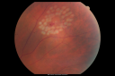
Retinal Tear After Laser1959 views65-year-old woman new onset floaters for 1 week. She initially saw flashing lights and then the flashing lights subsided and then she had the floaters VISUAL ACUITY: OD 20/25, OS 20/50
|
|

Multiple - Congenital Hypertrophy of the Pigment Epithelium - CHRPE - Gardner Syndrome1941 views63 Year Old woman with a history of colon cancer at age 32. She has also had thyroid cancer. Two of her 6 siblings died of cancer, one of sarcoma and one of lung cancer. Vision 20/30, 20/25. Fundus shows CHRPE in both eyes.
|
|

Progressive Outer Retinal Necrosis 77 Year Old Woman with CLL (Acute Retinal Necrosis) (PORN - ARN)1940 views77-year-old woman with CLL who had shingles on the left side of her face about 6 weeks ago then she developed a dendrite in the cornea which was treating about four weeks ago. She noticed severe vision loss in the left eye just a few days ago and you saw retinitis and she comes in because of that. Vision OD is 20/25, OS is hand motion
|
|

Plaquenil Toxicity both Eyes - Partial Bull's Eye - Discontinued 6 Years ago Fundus Autofluorescence1938 views82-year-old woman was on Plaquenil from 1976 from 2005, 200 mg a day. It was discontinued because of abnormal visual fields 6 years ago. OD 20/32, OS 20/40
|
|

Stargardt's Juvenille Macular Dystrophy - Fundus Flavimaculatus - Famlial1928 views55-year-old woman was seen in the office on 11/18/08. She has a half sister with Stargardt’s disease and seven other siblings who are fine. 20/120 OD, 20/160 OS.
|
|

Peripheral reticular pigment degeneration - pavingstone degeneration - age-related macular degeneration1924 views60 year old healthy man with intermediate AMD and peripheral pigment degeneration and pavinstone degeneration. Vision is 20/20 OU and the patient is asymptomatic. No significant history of AMD and he is a non-smoker.
|
|

Myelinated Nerve Fiber Layer Right Eye (white area)1915 views62-year-old man myelinated nerve fiber layer in the right eye. OD 20/25, OS 20/30.
|
|
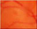
Acute Posterior Vitreous Detachment - Weiss Ring - Vogt Ring1910 views
|
|
| 17965 files on 180 page(s) |
1 |
 |
|
|
|
|
|
|
|
|
|


