|
|

OD Albinism with foveal hypoplasia2105 x angesehen28 year old female latina with vision of 20/400    
(6 Bewertungen)
|
|

Unilateral Hemorrhagic Retinopathy1194 x angesehenA 53 yr-old male was referred for a Macular Aneurysm. The FA should no apparent Aneurysm, nor did it show any evident VO or Edema. The patient is in the Navy but hadn't gone on any deep sea dives or any air flights.
The cause of the hemorrhages is unknown, the following ARVO abstract from 2012 identifies similar cases found in females.
Clinical Features Of Unilateral Hemorrhagic Retinopathy: A New Retinal Entity?
Presentation Start/End Time: Wednesday, May 09, 2012, 1:45 PM - 3:30 PM
Session 466    
(4 Bewertungen)
|
|

Macular fold after PPV for RD2698 x angesehenA 53-year-old man with a macula-off RD underwent left eye pars plana vitrectomy with air-fluid exchange, laser retinopexy, and injection of 14% SF6 gas. There were 4 retinal breaks between 10 o’clock and 12 o’clock including one large tear. He was compliant with facedown positioning. On the 8th post-operative day, a retinal fold along the 4-10 o’clock meridian was seen coursing through his central macula. He underwent repeat PPV but multiple attempts to lift and flatten the retina were unsuccessful.    
(4 Bewertungen)
|
|

Retinoblastoma1867 x angesehenYoung male who has a retinal tumor inferior with a calcium core in the left eye. This is a regressed retinoblastoma.     
(3 Bewertungen)
|
|

Reoccurring Retinal Detachment948 x angesehenPatient comes in for second opinion for RD in the right eye. Patient's VA was count fingers @ 2-ft in the right eye and 20/20 in the left eye. Patient is aphakic and has had 5- retinal surgeries in the past in the right eye including a scleral buckle. RD present with the macula off. Will consider surgery with silicone oil.     
(3 Bewertungen)
|
|

Rubeosis Iridis1230 x angesehenPatient presents with Rubeosis Iridis in the right eye due to neovascular glaucoma. VA is 20/40 in the right eye. Will follow up in 3-months.    
(3 Bewertungen)
|
|

Retinal Detachment with Dislocated IOL Lens1360 x angesehen47-year old male who had trauma to the right eye. Patient had retinal detachment surgery in the past (Scleral Buckle), to the right eye. Patient came in with another retinal detachment with dislocated PC IOL lens. Notice the haptics tearing the retina. Patient underwent vitrectomy with gas exchange. VA was hand motion 1-day post op.    
(3 Bewertungen)
|
|

Chronic Retinal Detachment936 x angesehen51 year old female, with a chronic retinal detachment. The patient has been stable since 1999.    
(3 Bewertungen)
|
|

Wyburn-Mason Syndrome 1893 x angesehen28 year old female, diagnosed with Wyburn-Mason Syndrome at age 7. At time of exam, vision in the left eye was 4/200.    
(3 Bewertungen)
|
|

Red Free Hemangioma left eye1738 x angesehen    
(3 Bewertungen)
|
|

Metastatic Choroidal Lesion986 x angesehenYoung female with choroidal lesion with subretinal fluid in the left eye. VA is finger count in the left eye.     
(2 Bewertungen)
|
|

Hypertrophy of the Retinal Pigment Epithelium1322 x angesehenPatient comes in with vision changes in the right eye. VA is 20/30 in the right eye. Fundus and FAF photos show a large area of Hypertrophy in the right eye.    
(2 Bewertungen)
|
|

Dislocated IOL with Tube Shunt1035 x angesehenPatient comes in with a dislocated lens in the right eye. Decreased vision in the right eye. Slit lamp photos shows lens dislocated halfway inferior. Ahmed Tube shut is visible at 11-o'clock superiorly in the right eye.     
(2 Bewertungen)
|
|

Gonioscopy, Mass in the Angle of Anterior Chamber1017 x angesehenSlit Lamp and goinoscopy photos show a mass at 7-o'clock i the right eye. The mass extends beneath the iris behind the lens.     
(2 Bewertungen)
|
|

Macular Scar1650 x angesehenFundus photography with Auto Fluorescent shows macular scar centrally, right eye. VA is 20/200 in the right eye.     
(2 Bewertungen)
|
|

Retinitis Pigmentosa1607 x angesehenPatient comes in with cloudy vision. VA is 20/200 in both eyes. Patient has had retinal pigmentosa since 1986. Patient to consider cataract surgery to help her visual symptoms...    
(2 Bewertungen)
|
|

Severe Pseudoexfoliation Ring, Left Eye843 x angesehenPatient comes in for blurred vision. VA is 20/30 both eyes. Mostly at near which was corrected with refraction. Slit lamp photos show severe pseudoexfoliation in the left eye. Patient will be followed up for glaucoma eval in a few weeks.     
(2 Bewertungen)
|
|

Corneal Foreign Body 1127 x angesehenPatient complains of foreign body sensation in the right eye. Slit lamp photos shows a piece of glass metal embedded into the inferior part of the cornea at 7-o'clock in the right eye. Foreign body was removed.     
(2 Bewertungen)
|
|

Ahmed Tube Placement1111 x angesehenPatient with severe glaucoma is seen post op for ahmed tube placement. VA is 20/70 and IOP is 8 in the left eye. Patient is stable.    
(2 Bewertungen)
|
|

Foreign Body "Rust Ring" 1213 x angesehenPatient complained of redness and tearing with white discharge. Patient was working with a grinding wheel on a vehicle. Slit lamp exam shows a rust ring barley off center. FBS was removed with Algiers Brush.    
(2 Bewertungen)
|
|
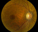
Full Thickness Macular Hole961 x angesehenFull Thickness Macular Hole    
(2 Bewertungen)
|
|

Bilateral Macular Star1201 x angesehenYoung female patient comes in with blurred vision in both eyes. VA is 20/40 in both eyes. Fundus photos show visible macular star centrally in both eyes. This is a result of Bilateral Neuroretinitis due to cat scratch.    
(2 Bewertungen)
|
|

Lupus Retinopathy985 x angesehenFemale patient comes in for eval on Lupus Retinopathy. Has poor vision in the right eye. VA is hand motion in the right eye. Fundus photos show fibrosis along the temporal arcades and narrowing of the arteries. No macular edema found.    
(2 Bewertungen)
|
|

Proliferative Diabetic Retinopathy1188 x angesehenFundus photography shows severe fibrosis and arterial narrowing. Peripheral laser scars in both eyes. VA is 20/40, right eye and 20/50, left eye.     
(2 Bewertungen)
|
|

Diabetic Retinopathy with CME1101 x angesehenSecond opinion for diabetic retinopathy. VA is 20/30, right eye, 20/50, left eye. Patient has mild swelling in the macula of the right eye. No evidence of CME but does have a moderate cataract in the left eye.     
(2 Bewertungen)
|
|

Giant Papillary Conjunctivitis911 x angesehenPatient wears soft contact lenses complained of irritation when the SCL would move. Inverted eyelid in both eyes and there was papillary +2 underneath the eyelid.     
(2 Bewertungen)
|
|

Subluxed IOL with TID1011 x angesehenPatient comes in with blurred vision in the left eye. Slit lamp exam shows dislocated IOL, inferior, left eye. PXF material on the implant inferiorly.    
(2 Bewertungen)
|
|

Retinal Detachment with Retinal Tear873 x angesehenPatient comes in with complaints of floaters in the right eye. VA was 20/40 with no improvement. Fundus exam shows retinal detachment from 9-12 o'clock with hole at 10:30 posteriorly. Pneumatic Retinopexy was performed with C3F8 Gas bubble and laser around the retinal tear in the right eye.     
(2 Bewertungen)
|
|

Choroidal Melanoma994 x angesehenPatient comes in with spot in vision in the right eye. Fundus photography shows medium size melanoma adjacent to the optic nerve and measures 13mm across horizontally and 6mm diameter in thickness. Patient will undergo proton beam therapy.     
(2 Bewertungen)
|
|

Bullseye Maculopathy without Plaquinel976 x angesehenFundus photograph reveals Bull's-eye maculopathy without the use of plaquinel. Patients VA is 20/20, Right eye and 20/25, Left eye. There are reported cases of Scleroderma patients with retinal pigment epithelial atrophy. Will return for follow up in 6-months.     
(2 Bewertungen)
|
|

Bullseye Maculopathy without Plaquinel994 x angesehenFundus photograph reveals Bull's-eye maculopathy without the use of plaquinel. Patients VA is 20/20, Right eye and 20/25, Left eye. There are reported cases of Scleroderma patients with retinal pigment epithelial atrophy. Will return for follow up in 6-months.     
(2 Bewertungen)
|
|
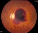
Retinal Hemorrhage 1186 x angesehenFundus photography shows central retinal hemorrhage. Patient was treated with Avastin Injection and will be followed up in 4-weeks.    
(2 Bewertungen)
|
|

Fibrosis Traction977 x angesehenYoung female patient with severe diabetes. Patients VA is 20/400 in the right eye. Fundus photos show a lot of fibrosis traction throughout the macula area. Fovea seems flat. Patient will undergo surgery for peeling of traction.     
(2 Bewertungen)
|
|
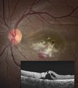
Cytomegalovirus Retinitis989 x angesehenPatient who is HIV Positive with Cytomegalovirus Retinitis. Patient was treated with Gangcyclovir Intra-Vitreal injection.    
(2 Bewertungen)
|
|

Choroidal Melanoma with Radiation Retinopathy1132 x angesehenPatient comes with follow up on Choroidal Melanoma, Right Eye that was treated back in June of 2009 with Radioactive Implant. Vein Occlusion is also present with VA - Hand Motion. Hemorrhages visible with hard exudates from the Radiation Retinopathy.     
(2 Bewertungen)
|
|

Cavernous Hemangioma967 x angesehen    
(2 Bewertungen)
|
|

Central Serous Retinopathy955 x angesehenYoung male who comes in for follow up on CSR in the left eye. Patient had focal laser treatment 1-month ago. There are still spots of leaking and proceeded with another focal laser treatment. Notice the top of the hyperfluorescent area.    
(2 Bewertungen)
|
|
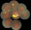
coats disease; exudative retinitis; retinal telangiectasis; leber multiple miliary aneurysm disease1722 x angesehenclassic coats    
(2 Bewertungen)
|
|

arteriovenous phase, acute recurrent CSR 655 x angesehenAngiogram focused on disc illustrates anterior displacement of the macula. Smokestack sign doesn't get any better than this.    
(2 Bewertungen)
|
|

fellow eye with subclinical areas of leakage586 x angesehen    
(2 Bewertungen)
|
|

PXE with Angioid Streaks1056 x angesehenPXE with angioid streaks OS (12/09/2011)    
(2 Bewertungen)
|
|

Endophthalmitis with Hypopion1235 x angesehen    
(2 Bewertungen)
|
|

HTN Retinopathy w/ cotton wool spots and hemorrhages1056 x angesehen23 year old african american male w/ visual acuity of 20/20    
(2 Bewertungen)
|
|

Horseshoe Retinal Tear with bridging blood vessel2420 x angesehen    
(2 Bewertungen)
|
|

Choroidal Melanoma1415 x angesehen    
(2 Bewertungen)
|
|

Choroidal Melanoma591 x angesehenChoroidal Melanoma with Retinal Detachment present inferior.    
(2 Bewertungen)
|
|

CNVM389 x angesehenPossible Chorioretinitis    
(2 Bewertungen)
|
|

BRVO666 x angesehen42 year old female VA 20/25
Branch Retinal Vein Occlusion - Hot Spot on Vein at Occlusion Site    
(2 Bewertungen)
|
|

MEWDS1009 x angesehenMultiple evanescent white dot syndrome (MEWDS) with classic "wreath pattern" and early optic nerve hyper fluorescence during the arterial phase of the fluorescein angiogram.     
(2 Bewertungen)
|
|

wyburn-mason syndrome fundus photos1105 x angesehen28 year old female, visual acuity 20/200    
(2 Bewertungen)
|
|

Solar retinopathy693 x angesehenA 27 year old African American male presented with a complaint of poor vision in both eyes for several months.
PMH: none
Medications: none
BCVA 20/40 OU
Slit-lamp exam: normal OU
On further questioning, he enjoys staring at the sun for 3-4 hours each day.    
(2 Bewertungen)
|
|

Central Retinal Artery Occlusion less than 24 hours - 69 year old man VA light perception615 x angesehen    
(2 Bewertungen)
|
|

Retinocytoma - Retinoma - Regressed Retinoblastoma 43 year old male incidental finding 1263 x angesehen    
(2 Bewertungen)
|
|

Optic Nerve Head Drusen Right Eye1568 x angesehen57-year-old woman has optic nerve drusen in both eyes.
VISUAL ACUITY: OU 20/20.     
(2 Bewertungen)
|
|
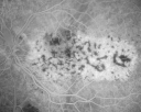
Bilateral Diffuse Uveal Melanocytic Proliferation - BDUMP - Paraneoplastic Syndrome1060 x angesehen80-year-old man vision loss for one year. He died about one year after these photos from Metastatic Poorly Differentiated Large Cell Carcinoma of unknown primary. He was a smoker.     
(2 Bewertungen)
|
|

Juxtapapillary CNVM and Serous Macular Detachment Wet AMD Rx Laser751 x angesehen85-year-old man OD is 20/25, OS is 20/100. IOP: OD 21, OS 22.
OS: Vertical C/D ratio is 0.1. There is a posterior vitreous separation. There is a hemorrhagic pigment epithelial detachment on the superior pole of the optic nerve, extending a disc-and-a-half diameter off the nerve, with adjacent exudates. There is a serous macular detachment involving the center of the fovea.
VA improved from 20/100 to 20/50 in four months from laser.    
(2 Bewertungen)
|
|

Free Floating Dislocated Lens in Vitreous978 x angesehenPatient comes in aphakic with dislocated lens floating to the back of the eye when laying down. Lens is laying up against the endothelium of the cornea when patient is right side up..    
(1 Bewertungen)
|
|

Flower Cataract1310 x angesehenPatient with ahmed tube and flower cataract    
(1 Bewertungen)
|
|
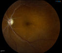
Retinal Artery Occlusion1447 x angesehenPatient comes in for eval on artery occlusions in both eyes. VA is 20/400, right eye and NLP, left eye. Fundus photos show paniretinal scars in the right eye with arterial narrowing and the left eye has arterial narrowing as well.    
(1 Bewertungen)
|
|

Vitreo-Traction due to Diabetic Retinopathy1093 x angesehenPatient comes in for follow up on her diabetes. VA is 20/30 in the right eye. Fundus photos shows thick traction nasally to the optic nerve forming a ring around the macula. Will follow up in 6-months.    
(1 Bewertungen)
|
|

Macular Pucker1112 x angesehenPatient comes in for followup on macular pucker, right eye. VA is 20/50 in the right eye...Fundus photo shows prominent pucker centrally pulling temporally in the right eye.    
(1 Bewertungen)
|
|

Retinal Tear with Laser Treatment1127 x angesehenPatient comes in with blurry vision and mild floaters in the left eye. Fundus photo shows retinal tear with fluid present in the superior nasal quadrant of the left eye. Laser Retinopexy was performed to fix the retinal tear.     
(1 Bewertungen)
|
|
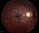
Acute Multifocal Placoid Pigment Epitheliopathy (AMPPE) related scarring1141 x angesehen    
(1 Bewertungen)
|
|
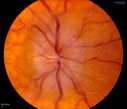
Bilateral Papilledema1147 x angesehenYoung female that presents with severe frequent headaches. VA is 20/15 in both eyes. Fundus exam reveals swelling of both optic nerves.    
(1 Bewertungen)
|
|

Bilateral Papilledema 1268 x angesehenYoung male presents with decreased vision in both eyes. Fundus photos reveal swollen optic nerves in both eyes.    
(1 Bewertungen)
|
|
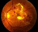
Nonproliferative Diabetic Retinopathy with Circinate Hard Exudates1325 x angesehen    
(1 Bewertungen)
|
|

Choriodal Rupture with Hemorrhage945 x angesehen    
(1 Bewertungen)
|
|

Silicone Oil Bubbles in the Angle1207 x angesehenPatient is evaluated for glaucoma associated with ocular inflammation. VA is CF in the right eye and 20/50, left eye. Gonioscopy photos shows little silicone oil bubbles in the inferior angle of the right eye. Patient to consider removal of silicone oil.     
(1 Bewertungen)
|
|

360-Degree Scleral Buckle1578 x angesehenPatient comes in with double vision from recent retinal detachment surgery. Optos shows 360 degree scleral buckle with retina attached. No holes or breaks visible. Patient had fresnel prism added on to lens to correct the double vision.    
(1 Bewertungen)
|
|

Bullous Retinal Detachment928 x angesehenPatient comes in with a curtain down in his vision in the right eye. Patient was hand motion. Fundus photograph shows retinal detachment with macula detached temporal superiorly in the right eye. There were 3-retinal tears between 10-12-o'clock. Schedule scleral buckle to fix RD.    
(1 Bewertungen)
|
|

Peripheral Iridotomy884 x angesehenSlit lamp photo of peripheral iridotomy for narrow angles. A hole was place at 10-o'clock superior temporally.     
(1 Bewertungen)
|
|

Treated Choroidal Melanoma851 x angesehenFollow up patient for melanoma in the right eye that was treated with proton beam therapy. VA is 20/50 in the right eye. Fundus photo shows scattered hemorrhages inferiorly with tumor temporal inferior at 8-o'clock. Patient will be followed up in 6-months    
(1 Bewertungen)
|
|

Melanoma of Optic Nerve979 x angesehenPatient comes in for evaluation on Optic Nerve Melanoma in the right eye. Patient's VA is 20/40 in the right eye. Fundus photograph reveals melanoma invading optic nerve and ultrasound reveals behind the optic nerve as well.     
(1 Bewertungen)
|
|

Dusted Pigment, Iris Nevus1163 x angesehen    
(1 Bewertungen)
|
|

Retinal Tear Surrounded By Laser Border856 x angesehen    
(1 Bewertungen)
|
|

Pterygium and RK Scars943 x angesehenPatient comes in for SCL fitting. VA is 20/30. right eye and 20/70, left eye. Slit lamp photos show 8-RK Scars in both eyes. Pterygium, nasally, left eye.     
(1 Bewertungen)
|
|

Morning Glory Syndrome, Optic Nerve1358 x angesehenPatient comes in for cataract evaluation. VA 20/60, right eye and 20/150, left eye. Fundus photo shows "morning glory syndrome" of the optic nerve in the left eye.     
(1 Bewertungen)
|
|
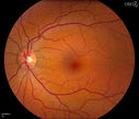
Pseudoxanthoma Elasticum with Angioid Streaks1015 x angesehenYoung female patient who comes in for routine eye exam, VA 20/20 in both eyes. Fundus photos shows a Peu'd orange appearance in the periphery of the retina and angioid streaks around the optic nerve.     
(1 Bewertungen)
|
|

Large Retinal Detachment with PVR1129 x angesehenPatient with poor vision in the left eye. Fundus photography shows rhegamatogenous retinal detachment with proliferative vitreoretinopathy in the left eye. Silicone oil is also present in the left eye.     
(1 Bewertungen)
|
|

Central Retinal Vein Occlusion with Disc Collaterals1012 x angesehen    
(1 Bewertungen)
|
|

Epiretinal Membrane with Flecks of Hemorrhage1090 x angesehen    
(1 Bewertungen)
|
|

Treated Melanoma784 x angesehenTreated melanoma in the right eye. VA is light perception in the right eye.     
(1 Bewertungen)
|
|
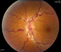
Stasis Retinopathy, Central Retinal Vein Occlusion1001 x angesehenYoung male who complains of decreased vision in the left eye. VA is 20/20 in the left eye. Fundus photo shows mild central retinal vein occlusion in the left eye.     
(1 Bewertungen)
|
|

Diabetic Retinopathy1239 x angesehenAnnual diabetic exam. VA 20/30, right eye and 20/50, left eye. Fundus photos shows hemorrhages with cotton wool spots. Exudates are visible around the macula in the right eye. Scattered hemorrhages in the left eye.    
(1 Bewertungen)
|
|

Epiretinal Membrane851 x angesehenPatient with a history of pars planitis comes in with decreased vision in the left eye. VA is 20/30 in the left eye. Fundus photos show a rather large epiretinal membrane covering the optic nerve in the left eye.     
(1 Bewertungen)
|
|

Central Retinal Vein Occlusion1129 x angesehenPatient comes in with mild blurred vision in the right eye. Fundus exam shows CRVO with scattered retinal hemorrhages in the right eye.    
(1 Bewertungen)
|
|

Choroidal Osteoma1181 x angesehenPatient with decreased vision within the past 2-years. VA 20/100 in the right eye. Fundus photo shows atrophic scar temporal to the macula in the right eye.    
(1 Bewertungen)
|
|

AVM948 x angesehenFP of AVM in OS of a 48 yr old female.    
(1 Bewertungen)
|
|

Optic Nerve Edema with Retinal Hemorrhages.839 x angesehenFemale patient diagnosed with lupus a year ago, comes in with spot and decreased vision in the right eye. VA is 20/400 in the right eye and 20/40, in the left eye. Fundus photography shows multiple retinal hemorrhages and possible bilateral papilledema.     
(1 Bewertungen)
|
|

Pseudoexfoliation Material Peeling.713 x angesehenPatient comes in for regular eye exam. Last exam was 8-years ago. Complains of decrease in VA. VA was 20/30, right eye and 20/40, left eye. Slit lamp photography shows pseudoexfoliation peeling of the lens within 360-degrees. Pseudoexfoliation ring is present centrally. IOP was normal. Will revisit in 3-months for follow up.    
(1 Bewertungen)
|
|

Intraocular Self injection839 x angesehenThe treatment of wet macular degeneration was revolutionized in late 2013 when Geneneron introduced its innovative and inexpensive take-home kit. Now, millions of patients around the world treat themselves on a PRN basis, using instructions written in 10 different languages.
April Fool!    
(1 Bewertungen)
|
|

Traumatic Chorioretinal Scar629 x angesehen    
(1 Bewertungen)
|
|
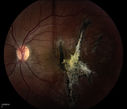
Choroidal Rupture Secondary to Ocular Trauma807 x angesehen    
(1 Bewertungen)
|
|
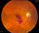
Branch Retinal Vein Occlusion924 x angesehenPatient comes in for eval on retinal hemorrhage in the right eye. VA is 20/30 in the right eye. Fundus photography shows branch retinal vein occlusion inferior of the macula with retinal hemorrhage present. Will reevaluate in 2-months.    
(1 Bewertungen)
|
|

Retinal Detachment with Macula Detached871 x angesehenPatient had sudden loss of vision in the left eye. VA is 20/60 in the left eye. Fundus exam reveals retinal detachment inferiorly from 3-9 o'clock with tear at 3:30. Patient underwent scleral buckle in the left eye.    
(1 Bewertungen)
|
|

Choroidal Nevus956 x angesehenPatient comes in with pigmented spot in the right eye. VA is 20/25. Fundus photography shows elevated choroidal nevus inferiorily in the right eye. Will be reevaluated in 3-months...     
(1 Bewertungen)
|
|

Choroidal Melanoma927 x angesehenFemale patient comes in with a visual defect in the right eye. VA is 20/40 in the right eye. Fundus photography shows choroidal melanoma with retinal detachment temporally at 3-o'clock in the right eye.    
(1 Bewertungen)
|
|

Choroidal Melanoma with Sub Retinal Fluid982 x angesehenFundus photography shows choroidal melanoma superiorily between 11-12-o'clock in the left eye. VA is 20/150 in the left eye.     
(1 Bewertungen)
|
|

Macular Hemorrhage911 x angesehenPatient comes in with central vision loss in the right eye. Fundus photography shows a macular hemorrhage in the central portion of the fovea. Cotton wool spots are present around the optic nerve and a little hemorrhage present inferiorily at 6-o'clock.    
(1 Bewertungen)
|
|

Neovascularization, Rubiosis870 x angesehenPatient comes in for GPCL fitting. VA is 20/50 w/GPCL. Slit lamp photography shows neovascularization on the iris in the right eye. Also iris atrophy, superiorly in the right eye.    
(1 Bewertungen)
|
|
| 454 Dateien auf 5 Seite(n) |
1 |
 |
 |
|